1
views
0
recommends
+1 Recommend
0 collections
    0
    shares
      • Record: found
      • Abstract: found
      • Article: found
      Is Open Access

      Multi-center clinical study using optical coherence tomography for evaluation of cervical lesions in-vivo

      research-article

      Read this article at

      Bookmark
          There is no author summary for this article yet. Authors can add summaries to their articles on ScienceOpen to make them more accessible to a non-specialist audience.

          Abstract

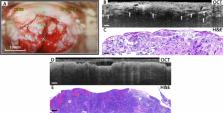
          In this prospective study of an in-vivo cervical examination using optical coherence tomography (OCT), we evaluated the diagnostic value of non-invasive and real-time OCT in cervical precancerous lesions and cancer diagnosis, and determined the characteristics of OCT images. 733 patients from 5 Chinese hospitals were inspected with OCT and colposcopy-directed biopsy. The OCT images were compared with the histological sections to find out the characteristics of various categories of lesions. The OCT images were also interpreted by 3 investigators to make a 2-class classification, and the results were compared against the pathological results. Various structures of the cervical tissue were clearly observed in OCT images, which matched well with the corresponding histological sections. The OCT diagnosis results delivered a sensitivity of 87.0% (95% confidence interval, CI 82.2–90.7%), a specificity of 84.1% (95% CI 80.3–87.2%), and an overall accuracy of 85.1%. Both good consistency of OCT images and histological images and satisfactory diagnosis results were provided by OCT. Due to its features of non-invasion, real-time, and accuracy, OCT is valuable for the in-vivo evaluation of cervical lesions and has the potential to be one of the routine cervical diagnosis methods.

          Related collections

          Most cited references22

          • Record: found
          • Abstract: found
          • Article: not found

          Global Cancer Statistics 2018: GLOBOCAN Estimates of Incidence and Mortality Worldwide for 36 Cancers in 185 Countries

          This article provides a status report on the global burden of cancer worldwide using the GLOBOCAN 2018 estimates of cancer incidence and mortality produced by the International Agency for Research on Cancer, with a focus on geographic variability across 20 world regions. There will be an estimated 18.1 million new cancer cases (17.0 million excluding nonmelanoma skin cancer) and 9.6 million cancer deaths (9.5 million excluding nonmelanoma skin cancer) in 2018. In both sexes combined, lung cancer is the most commonly diagnosed cancer (11.6% of the total cases) and the leading cause of cancer death (18.4% of the total cancer deaths), closely followed by female breast cancer (11.6%), prostate cancer (7.1%), and colorectal cancer (6.1%) for incidence and colorectal cancer (9.2%), stomach cancer (8.2%), and liver cancer (8.2%) for mortality. Lung cancer is the most frequent cancer and the leading cause of cancer death among males, followed by prostate and colorectal cancer (for incidence) and liver and stomach cancer (for mortality). Among females, breast cancer is the most commonly diagnosed cancer and the leading cause of cancer death, followed by colorectal and lung cancer (for incidence), and vice versa (for mortality); cervical cancer ranks fourth for both incidence and mortality. The most frequently diagnosed cancer and the leading cause of cancer death, however, substantially vary across countries and within each country depending on the degree of economic development and associated social and life style factors. It is noteworthy that high-quality cancer registry data, the basis for planning and implementing evidence-based cancer control programs, are not available in most low- and middle-income countries. The Global Initiative for Cancer Registry Development is an international partnership that supports better estimation, as well as the collection and use of local data, to prioritize and evaluate national cancer control efforts. CA: A Cancer Journal for Clinicians 2018;0:1-31. © 2018 American Cancer Society.
            Bookmark
            • Record: found
            • Abstract: not found
            • Article: not found

            Optical coherence tomography

              Bookmark
              • Record: found
              • Abstract: found
              • Article: not found

              Two-sided confidence intervals for the single proportion: comparison of seven methods.

              Simple interval estimate methods for proportions exhibit poor coverage and can produce evidently inappropriate intervals. Criteria appropriate to the evaluation of various proposed methods include: closeness of the achieved coverage probability to its nominal value; whether intervals are located too close to or too distant from the middle of the scale; expected interval width; avoidance of aberrations such as limits outside [0,1] or zero width intervals; and ease of use, whether by tables, software or formulae. Seven methods for the single proportion are evaluated on 96,000 parameter space points. Intervals based on tail areas and the simpler score methods are recommended for use. In each case, methods are available that aim to align either the minimum or the mean coverage with the nominal 1 -alpha.
                Bookmark

                Author and article information

                Contributors
                zxa@zzu.edu.cn
                307797362@qq.com
                Journal
                Sci Rep
                Sci Rep
                Scientific Reports
                Nature Publishing Group UK (London )
                2045-2322
                5 April 2021
                5 April 2021
                2021
                : 11
                : 7507
                Affiliations
                [1 ]GRID grid.412719.8, Department of Gynecology, , The Third Affiliated Hospital of Zhengzhou University, ; Zhengzhou, 450052 China
                [2 ]GRID grid.412719.8, Department of Pathology, , The Third Affiliated Hospital of Zhengzhou University, ; Zhengzhou, 450052 China
                [3 ]GRID grid.459742.9, ISNI 0000 0004 1798 5889, Department of Gynecology, , Liaoning Cancer Hospital and Institute, ; Shenyang, 110042 China
                [4 ]Department of Gynecology, Luohe Central Hospital, Luohe, 462000 China
                [5 ]Department of Gynecology, Puyang Oilfield General Hospital, Puyang, 457001 China
                [6 ]GRID grid.508190.5, Department of Gynecology, , Zhengzhou Jinshui District General Hospital, ; Zhengzhou, 450002 China
                [7 ]Zhengzhou Ultralucia Medical Technology Ltd., Zhengzhou, 450016 China
                [8 ]GRID grid.412719.8, Department of Imaging, , The Third Affiliated Hospital of Zhengzhou University, ; Zhengzhou, 450052 China
                Article
                86711
                10.1038/s41598-021-86711-3
                8021566
                33820924
                3541f2c9-5a78-4ce0-9b05-d1601347dc37
                © The Author(s) 2021

                Open Access This article is licensed under a Creative Commons Attribution 4.0 International License, which permits use, sharing, adaptation, distribution and reproduction in any medium or format, as long as you give appropriate credit to the original author(s) and the source, provide a link to the Creative Commons licence, and indicate if changes were made. The images or other third party material in this article are included in the article's Creative Commons licence, unless indicated otherwise in a credit line to the material. If material is not included in the article's Creative Commons licence and your intended use is not permitted by statutory regulation or exceeds the permitted use, you will need to obtain permission directly from the copyright holder. To view a copy of this licence, visit http://creativecommons.org/licenses/by/4.0/.

                History
                : 8 May 2020
                : 13 January 2021
                Categories
                Article
                Custom metadata
                © The Author(s) 2021

                Uncategorized
                diseases,medical research,oncology
                Uncategorized
                diseases, medical research, oncology

                Comments

                Comment on this article