9
views
0
recommends
+1 Recommend
0 collections
    0
    shares
      • Record: found
      • Abstract: found
      • Article: found
      Is Open Access

      Leishmania donovani development in Phlebotomus argentipes: comparison of promastigote- and amastigote-initiated infections

      research-article

      Read this article at

      Bookmark
          There is no author summary for this article yet. Authors can add summaries to their articles on ScienceOpen to make them more accessible to a non-specialist audience.

          SUMMARY

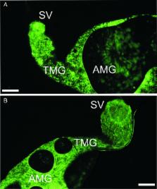
          Leishmania parasites alternate in their life cycle between promastigote stages that develop in the gut of phlebotomine sand flies and amastigotes residing inside phagocytic cells of vertebrate hosts. For experimental infections of sand flies, promastigotes are frequently used as this way of infection is technically easier although ingestion of promastigotes by sand flies is unnatural. Here we aimed to answer a critical question, to what extent do promastigote-initiated experimental infections differ from those initiated with intracellular amastigotes. We performed side-by-side comparison of Leishmania development in Phlebotomus argentipes females infected alternatively with promastigotes from log-phase cultures or amastigotes grown ex vivo in macrophages. Early stage infections showed substantial differences in parasite load and representation of morphological forms. The differences disappeared along the maturation of infections; both groups developed heavy late-stage infections with colonization of the stomodeal valve, uniform representation of infective metacyclics and equal efficiency of transmission. The results showed that studies focusing on early phase of Leishmania development in sand flies should be initiated with intracellular amastigotes. However, the use of promastigote stages for sand fly infections does not alter significantly the final outcome of Leishmania donovani development in P. argentipes and their transmissibility to the vertebrate host.

          Related collections

          Most cited references35

          • Record: found
          • Abstract: found
          • Article: not found

          Transmission of Leishmania metacyclic promastigotes by phlebotomine sand flies

          A thorough understanding of the transmission mechanism of any infectious agent is crucial to implementing an effective intervention strategy. Here, our current understanding of the mechanisms that Leishmania parasites use to ensure their transmission from sand fly vectors by bite is reviewed. The most important mechanism is the creation of a “blocked fly” resulting from the secretion of promastigote secretory gel (PSG) by the parasites in the anterior midgut. This forces the sand fly to regurgitate PSG before it can bloodfeed, thereby depositing both PSG and infective metacyclic promastigotes in the skin of a mammalian host. Other possible factors in transmission are considered: damage to the stomodeal valve; occurrence of parasites in the salivary glands; and excretion of parasites from the anus of infected sand flies. Differences in the transmission mechanisms employed by parasites in the three subgenera, Leishmania, Viannia and Sauroleishmania are also addressed.
            Bookmark
            • Record: found
            • Abstract: found
            • Article: found
            Is Open Access

            Leishmania development in sand flies: parasite-vector interactions overview

            Leishmaniases are vector-borne parasitic diseases with 0.9 – 1.4 million new human cases each year worldwide. In the vectorial part of the life-cycle, Leishmania development is confined to the digestive tract. During the first few days after blood feeding, natural barriers to Leishmania development include secreted proteolytic enzymes, the peritrophic matrix surrounding the ingested blood meal and sand fly immune reactions. As the blood digestion proceeds, parasites need to bind to the midgut epithelium to avoid being excreted with the blood remnant. This binding is strictly stage-dependent as it is a property of nectomonad and leptomonad forms only. While the attachment in specific vectors (P. papatasi, P. duboscqi and P. sergenti) involves lipophosphoglycan (LPG), this Leishmania molecule is not required for parasite attachment in other sand fly species experimentally permissive for various Leishmania. During late-stage infections, large numbers of parasites accumulate in the anterior midgut and produce filamentous proteophosphoglycan creating a gel-like plug physically obstructing the gut. The parasites attached to the stomodeal valve cause damage to the chitin lining and epithelial cells of the valve, interfering with its function and facilitating reflux of parasites from the midgut. Transformation to metacyclic stages highly infective for the vertebrate host is the other prerequisite for effective transmission. Here, we review the current state of knowledge of molecular interactions occurring in all these distinct phases of parasite colonization of the sand fly gut, highlighting recent discoveries in the field.
              Bookmark
              • Record: found
              • Abstract: found
              • Article: not found

              Quantification of Leishmania infantum DNA by a real-time PCR assay with high sensitivity.

              A real-time PCR was developed to quantify Leishmania infantum kinetoplast DNA and optimized to reach a sensitivity of 0.0125 parasites/ml of blood. In order to analyze the incidence of heterogeneity and number of minicircles, we performed comparative PCR by using the Leishmania DNA polymerase gene as a reporter. Assays performed in both promastigote and amastigote stages showed variations among different L. infantum and Leishmania donovani strains and the stability of the minicircle numbers for a particular strain. Analysis of blood samples from a patient who presented with Mediterranean visceral leishmaniasis confirmed the reliability of such an assay for Leishmania quantification in biological samples and allowed an estimation of positivity thresholds of classical tests used for direct diagnosis of the disease; positivity thresholds were in the range of 18 to 42, 0.7 to 42, and 0.12 to 22.5 parasites/ml for microscopic examination, culture, and conventional PCR, respectively. At the time of diagnosis, parasitemia could vary by a wide range (32 to 188,700 parasites/ml, with a median of 837 parasites/ml), while in bone marrow, parasite load was more than 100 parasites per 10(6) nucleated human cells. After successful therapy, parasitemia levels remain lower than 1 parasite/ml. In the immunocompromised host, relapses correlate with an increase in the level of parasitemia, sometimes scanty, justifying the need for assays with high sensitivity. Such sensitivity allows the detection of Leishmania DNA in the blood of 21% of patients with no history of leishmaniasis living in the Marseilles area, where leishmaniasis is endemic. This technique may be useful for epidemiologic and diagnostic purposes, especially for the quantification of parasitemia at low levels during posttherapy follow-up.
                Bookmark

                Author and article information

                Journal
                Parasitology
                Parasitology
                PAR
                Parasitology
                Cambridge University Press (Cambridge, UK )
                0031-1820
                1469-8161
                April 2017
                23 November 2016
                : 144
                : 4
                : 403-410
                Affiliations
                [1 ]Department of Parasitology, Faculty of Science, Charles University , Vinicna 7, 128 44 Prague 2, Czech Republic
                [2 ]Department of Pathogen Molecular Biology, Faculty of Infectious and Tropical Diseases, London School of Hygiene and Tropical Medicine , Keppel Street, WC1E 7HTLondon, UK
                Author notes
                [* ]Corresponding author: Department of Parasitology, Faculty of Science, Charles University , Vinicna 7, 128 44 Prague 2, Czech Republic. E-mail: sadlovaj@ 123456natur.cuni.cz
                Article
                S0031182016002067 00206
                10.1017/S0031182016002067
                5368687
                27876097
                35083133-5f8c-4abe-bff9-d4f8725c0159
                © Cambridge University Press 2016

                This is an Open Access article, distributed under the terms of the Creative Commons Attribution licence ( http://creativecommons.org/licenses/by/4.0/), which permits unrestricted re-use, distribution, and reproduction in any medium, provided the original work is properly cited.

                History
                : 02 August 2016
                : 12 October 2016
                : 15 October 2016
                Page count
                Figures: 5, Tables: 1, References: 35, Pages: 8
                Categories
                Research Article

                Parasitology
                leishmania donovani,phlebotomus,promastigotes,amastigotes,transmission
                Parasitology
                leishmania donovani, phlebotomus, promastigotes, amastigotes, transmission

                Comments

                Comment on this article