26
views
0
recommends
+1 Recommend
0 collections
    0
    shares
      • Record: found
      • Abstract: found
      • Article: found
      Is Open Access

      EMPHYSEMATOUS PYELONEPHRITIS

      letter

      Read this article at

      Bookmark
          There is no author summary for this article yet. Authors can add summaries to their articles on ScienceOpen to make them more accessible to a non-specialist audience.

          Abstract

          Dear Editor, A 48-year-old female with poorly controlled diabetes mellitus (DM) presented herself with a five-day history of fever, chills, left flank pain and dysuria. On admission, her abdomen was mildly distended and tender over the left lumbar region. Laboratory data showed white blood cell count of 18.6×10⁹/L with 91.7% neutrophils, serum creatinine at 223 μmol/L, C-reactive protein at 168 mg/L and glycosylated hemoglobin (HbA1c) at 14.1%. Urine analysis showed turbid appearance with obvious pyuria, hematuria and proteinuria. A renal ultrasound scan revealed potential signs of gas in the parenchyma of the left kidney. A 
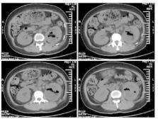
non-contrast computed tomography (CT) scan of the abdomen demonstrated swelling of the left kidney with visible gas in the renal parenchyma (Fig. 1), radiologically associated with emphysematous pyelonephritis (EPN, Class 2). The patient underwent CT-guided percutaneous catheter drainage (PCD) and was treated with broad-spectrum intravenous antibiotics and rigorous blood sugar control. The urine and pus cultures showed significant growth of Escherichia coli (E. coli). From the above medical procedures, the patient improved significantly and was discharged with an excellent prognosis. EPN is an uncommon, but acutely severe and life-threatening necrotizing kidney infection, which is characterized by gas accumulation in the renal parenchyma, collecting system, or perinephric tissue1 , 2 , 4. The disease usually occurs in female patients with poorly controlled DM, with or without urinary tract obstruction1 , 2 , 4. E. coli is the most common pathogen, which has been extracted from urine or pus cultures in almost 70% of the patients4. EPN is a radiological diagnosis, with CT scan currently being the imaging procedure of choice for early diagnosis and assessment of the disease1 , 2 , 4. Importantly, PCD is now the most appropriate strategy and the gold standard in management of EPN2 , 3. Over the last two decades, improvements in management techniques have drastically reduced the mortality rate of EPN to 21%3 , 4. Fig. 1 Computed tomography (CT) scan of the abdomen showing a mottled gas collection within the parenchyma of the swelling left kidney

          Related collections

          Most cited references5

          • Record: found
          • Abstract: found
          • Article: not found

          Emphysematous pyelonephritis: clinicoradiological classification, management, prognosis, and pathogenesis.

          Emphysematous pyelonephritis (EPN) is a rare, severe gas-forming infection of renal parenchyma and its surrounding areas. The radiological classification and adequate therapeutic regimen are controversial and the prognostic factors and pathogenesis remain uncertain. To elucidate the clinical features, radiological classification, and prognostic factors of EPN; to compare the modalities of management (ie, antibiotic treatment alone, percutaneous catheter drainage combined with antibiotic treatment, or nephrectomy) and outcome among the various radiological classes of EPN; and to clarify the gas-forming mechanism and pathogenesis of EPN by gas analysis and pathological findings. Forty-eight EPN cases from our institution were enrolled between August 1,1989, and November 30, 1997. According to the radiological findings on computed tomographic scan, they were classified into the following classes: (1) class 1: gas in the collecting system only; (2) class 2: gas in the renal parenchyma without extension to extrarenal space; (3) class 3A: extension of gas or abscess to perinephric space; class 3B: extension of gas or abscess to pararenal space; and (4) class 4: bilateral EPN or solitary kidney with EPN. The clinical manifestations, management, and outcome were compared. The gas contents of specimens from 6 patients were analyzed. The pathological findings from 8 patients who received nephrectomy were reviewed. The statistical methods consisted of the Fisher exact test (2 tailed) for categorical variables and Wilcoxon rank sum test for continuous variables to test the predictors of poor prognosis. Forty-six patients (96%) had diabetes mellitus, and 10 (22%) of the 46 also had urinary tract obstruction in the corresponding renoureteral unit. The other 2 nondiabetic patients (4%) had severe hydronephrosis. Twenty-one (72%) of the 29 patients with diabetes mellitus also had a glycosylated hemoglobin A(1c) level higher than 0.08. Escherichia coli (69%) and Klebsiella pneumoniae (29%) were the most common pathogens. The mortality rate in patients who received antibiotic treatment alone was 40% (2 of 5 patients). The success rate of management by percutaneous catheter drainage (PCD) combined with antibiotic treatment was 66% (27 of 41 patients). In classes 1 and 2 EPN, all the patients who were treated using a PCD or ureteral catheter combined with antibiotic treatment survived. In extensive EPN (classes 3 and 4), 17 (85%) of the 20 patients with fewer than 2 risk factors (ie, thrombocytopenia, acute renal function impairment, disturbance of consciousness, or shock) were successfully treated using PCD combined with antibiotic treatment; and the patients with 2 or more risk factors had a significantly higher failure rate than those with no or only 1 risk factors (92% vs 15%, P<.001). Eight of the 14 patients who had an unsuccessful treatment using a PCD underwent subsequent nephrectomy, 7 of whom survived. Only 2 patients were managed by direct nephrectomy and survived. The overall success rate of nephrectomy was 90% (9 of 10 patients). The total mortality was 18.8% (9 of 48 patients). Five of the 6 gas samples contained hydrogen (average, 12.8%), and all had carbon dioxide (average, 14.4%). The pathological findings from 8 of 10 who underwent nephrectomy revealed poor perfusion in most cases (ie, infarction, 5 patients; vascular thrombosis, 3 patients; and arteriosclerosis and/or glomerulosclerosis, 4 patients). Acute renal infection with E coli or K pneumoniae in patients with diabetes mellitus and/or urinary tract obstruction is the cornerstone for the development of EPN. Mixed acid fermentation of glucose by Enterobacteriaceae is the major pathway of gas formation. For localized EPN (classes 1 and 2), PCD combined with antibiotic treatment can provide a good outcome. (ABSTRACT TRUNCATED)
            Bookmark
            • Record: found
            • Abstract: found
            • Article: not found

            Emphysematous pyelonephritis.

            Emphysematous pyelonephritis (EPN) is a severe necrotizing infection of the renal parenchyma. The clinical course of EPN can be severe and life-threatening if not recognized and treated promptly. Most of the information has been from case reports, a few large series have also been reported. Using an evidence-based approach, this review describes the pathogenesis, classification, complications, and management of EPN. Emphysematous pyelonephritis (EPN) is an acute severe necrotizing infection of the renal parenchyma and its surrounding tissues that results in the presence of gas in the renal parenchyma, collecting system, or perinephric tissue. The cause for mortality in EPN is primarily due to septic complications. Up to 95% of the cases with EPN have underlying uncontrolled diabetes mellitus. The risk of developing EPN secondary to a urinary tract obstruction is about 25-40%. There are three classifications of EPN based on radiological findings. Acute renal failure, microscopic or macroscopic haematuria, severe proteinuria are other positive findings in EPN. Escherichia coli is the most common causative pathogen with the organism isolated on urine or pus cultures in nearly 70% of the reported cases. A plain radiograph shows an abnormal gas shadow in the renal bed raising the suspicion whereas an ultrasound scan or computed tomography (CT) will confirm the presence of intra-renal gas thus supporting the diagnosis of EPN. Gas may extend beyond the site of inflammation to the sub capsular, perinephric and pararenal spaces. In some cases, gas was found to be extending into the scrotal sac and spermatic cord. Subsequent case studies have shown patients being successfully treated with PCD when used in addition to medical management, with significant reduction in the morality rates. PCD should be performed on patients who have localized areas of gas and functioning renal tissue is present. The treatment strategies include MM alone, PCD plus MM, MM plus emergency nephrectomy, and PCD plus MM plus emergency nephrectomy. In small proportion of patients managed with MM and PCD, subsequent nephrectomy will be required and in these patients the reported mortality is 6.6% Nephrectomy in patients with EPN can be simple, radical or laparoscopic. © 2010 THE AUTHORS. BJU INTERNATIONAL © 2010 BJU INTERNATIONAL.
              Bookmark
              • Record: found
              • Abstract: found
              • Article: not found

              Is percutaneous drainage the new gold standard in the management of emphysematous pyelonephritis? Evidence from a systematic review.

              There is no current consensus on what constitutes the ideal management of emphysematous pyelonephritis. We review the current management strategies including the role of nephron preserving percutaneous drainage in the treatment of emphysematous pyelonephritis. We searched MEDLINE, PubMed, EMBASE, CINAHL and the Cochrane Library from 1966 to 2006. Abstracts were reviewed including all types of studies from prospective randomized controlled studies to small retrospective series. All relevant English language articles reporting on at least 5 patients were reviewed. Ten retrospective studies on 210 patients with emphysematous pyelonephritis met the inclusion criteria. There were 167 females and 43 males with a mean age of 57 years (range 24 to 83). Of the patients 96% had diabetes mellitus and 29% had urinary tract obstruction. The diagnostic accuracy of computerized tomography was 100%. Escherichia coli and Klebsiella were the most common causative agents. The mortality from medical management alone was 50%, medical management combined with emergency nephrectomy was 25% and medical management combined with percutaneous drainage was 13.5%. Mortality was significantly less in patients undergoing percutaneous drainage compared to other treatments (Pearson chi-square p <0.001). Of the patients who underwent medical treatment with percutaneous drainage a small number (15) underwent elective nephrectomy and mortality was 6.6% (1 of 15). Percutaneous drainage should be part of the initial management strategy for emphysematous pyelonephritis. This strategy is associated with a lower mortality than medical management or emergency nephrectomy. Delayed elective nephrectomy may be required in some patients.
                Bookmark

                Author and article information

                Journal
                Rev Inst Med Trop Sao Paulo
                Rev. Inst. Med. Trop. Sao Paulo
                rimtsp
                Revista do Instituto de Medicina Tropical de São Paulo
                Instituto de Medicina Tropical
                0036-4665
                1678-9946
                Jul-Aug 2015
                Jul-Aug 2015
                : 57
                : 4
                : 368
                Affiliations
                [1 ]originalDepartment of Infectious Diseases, Shanghai Jiao Tong University Affiliated Sixth People's Hospital, Shanghai, P. R. China normalizedShanghai Jiaotong University orgdiv2Department of Infectious Diseases orgnameShanghai Jiao Tong University orgdiv1Affiliated Sixth People's Hospital Shanghai P. R.China
                Author notes
                Correspondence to: Yong-Sheng YU, Department of Infectious Diseases, Shanghai Jiao Tong University Affiliated Sixth People's Hospital, 600 Yishan Road, 200233 Shanghai, P. R. China Phone: + 86 21 64369181-58673; Fax: + 86 21 24058384 E-mail: yuyongsheng@ 123456medmail.com.cn
                Article
                10.1590/S0036-46652015000400019
                4616928
                26422167
                34feb388-bf87-4c2d-9af3-fd9f7b3d5dc1

                This is an open-access article distributed under the terms of the Creative Commons Attribution License

                History
                Page count
                Figures: 1, Tables: 0, Equations: 0, References: 4, Pages: 1
                Funding
                Funded by: Shanghai Jiao Tong University School of Medicine
                Award ID: YB130910
                This work was partly supported by the Medical Education Research Project of Shanghai Jiao Tong University School of Medicine (No. YB130910)
                Categories
                Letter to the Editor

                Comments

                Comment on this article