12
views
0
recommends
+1 Recommend
0 collections
    0
    shares
      • Record: found
      • Abstract: found
      • Article: found
      Is Open Access

      Septins are critical regulators of osteoclastic bone resorption

      research-article

      Read this article at

      Bookmark
          There is no author summary for this article yet. Authors can add summaries to their articles on ScienceOpen to make them more accessible to a non-specialist audience.

          Abstract

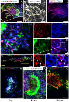
          Septins are known to play key roles in supporting cytoskeletal stability, vesicular transport, endo-/exocytosis, stabilizing cellular membranes and forming diffusion barriers. Their function in mammalian cells is poorly investigated. The osteoclast offers an interesting tool to investigate septins because all cellular activities septins were reported to be involved in are critical for osteoclasts. However, the existence of septins in osteoclasts has not even been reported. Here we show that the SEPT9 gene and Septin 9 (SEPT9) protein are expressed and synthesized during differentiation of human osteoclasts. Pharmacological stabilization of septin filaments dose dependently inhibits bone resorption of human osteoclasts in vitro suggesting a role for septins in bone resorption. Attesting to this, conditional deletion of Sept9 in mice leads to elevated levels of trabecular bone and diminished femoral growth in vivo. Finally, systematic interrogation of the spatial organization of SEPT9 by confocal microscopy reveals that SEPT9 is closely associated to the structures known to be critical for osteoclast activity. We propose that septins in general and SEPT9 in particular play a previously unappreciated role in osteoclastic bone resorption.

          Related collections

          Most cited references55

          • Record: found
          • Abstract: found
          • Article: not found

          A septin diffusion barrier at the base of the primary cilium maintains ciliary membrane protein distribution.

          In animal cells, the primary cilium transduces extracellular signals through signaling receptors localized in the ciliary membrane, but how these ciliary membrane proteins are retained in the cilium is unknown. We found that ciliary membrane proteins were highly mobile, but their diffusion was impeded at the base of the cilium by a diffusion barrier. Septin 2 (SEPT2), a member of the septin family of guanosine triphosphatases that form a diffusion barrier in budding yeast, localized at the base of the ciliary membrane. SEPT2 depletion resulted in loss of ciliary membrane protein localization and Sonic hedgehog signal transduction, and inhibited ciliogenesis. Thus, SEPT2 is part of a diffusion barrier at the base of the ciliary membrane and is essential for retaining receptor-signaling pathways in the primary cilium.
            Bookmark
            • Record: found
            • Abstract: found
            • Article: not found

            Self- and actin-templated assembly of Mammalian septins.

            Septins are polymerizing GTPases required for cytokinesis and cortical organization. The principles by which they are targeted to, and assemble at, specific cell regions are unknown. We show that septins in mammalian cells switch between a linear organization along actin bundles and cytoplasmic rings, approximately 0.6 microm in diameter. A recombinant septin complex self-assembles into rings resembling those in cells. Linear organization along actin bundles was reconstituted by adding an adaptor protein, anillin. Perturbation of septin organization in cells by expression of a septin-interacting fragment of anillin or by septin depletion via siRNA causes loss of actin bundles. We conclude that septins alone self-assemble into rings, that adaptor proteins recruit septins to actin bundles, and that septins help organize these bundles.
              Bookmark
              • Record: found
              • Abstract: not found
              • Article: not found

              Genetic control of the cell division cycle in yeast. IV. Genes controlling bud emergence and cytokinesis.

              S Hartwell (1971)
                Bookmark

                Author and article information

                Contributors
                kent.soee@rsyd.dk
                Journal
                Sci Rep
                Sci Rep
                Scientific Reports
                Nature Publishing Group UK (London )
                2045-2322
                29 August 2018
                29 August 2018
                2018
                : 8
                : 13016
                Affiliations
                [1 ]ISNI 0000 0001 0728 0170, GRID grid.10825.3e, Department of Clinical Cell Biology, Vejle Hospital/Lillebaelt Hospital, , Institute of Regional Health Research, University of Southern Denmark, ; Beriderbakken 4, 7100 Vejle, Denmark
                [2 ]ISNI 0000 0001 1956 2722, GRID grid.7048.b, Department of Molecular Biology and Genetics, , Aarhus University, ; 8000 Aarhus, Denmark
                [3 ]ISNI 0000 0001 1956 2722, GRID grid.7048.b, Department of Biomedicine, , Aarhus University, ; 8000 Aarhus, Denmark
                [4 ]ISNI 0000 0004 1936 7910, GRID grid.1012.2, Cellular Orthopaedic Laboratory, School of Biomedical Sciences, , University of Western Australia, ; Nedlands, 6009 Australia
                Author information
                http://orcid.org/0000-0002-9320-7195
                http://orcid.org/0000-0001-7402-314X
                Article
                31159
                10.1038/s41598-018-31159-1
                6115361
                30158637
                34293de1-4c5b-469c-8d6f-5fc3b0bb6b7b
                © The Author(s) 2018

                Open Access This article is licensed under a Creative Commons Attribution 4.0 International License, which permits use, sharing, adaptation, distribution and reproduction in any medium or format, as long as you give appropriate credit to the original author(s) and the source, provide a link to the Creative Commons license, and indicate if changes were made. The images or other third party material in this article are included in the article’s Creative Commons license, unless indicated otherwise in a credit line to the material. If material is not included in the article’s Creative Commons license and your intended use is not permitted by statutory regulation or exceeds the permitted use, you will need to obtain permission directly from the copyright holder. To view a copy of this license, visit http://creativecommons.org/licenses/by/4.0/.

                History
                : 20 June 2018
                : 7 August 2018
                Funding
                Funded by: This study was supported by general research funding from Lillebaelt Hospital and Aarhus University.
                Categories
                Article
                Custom metadata
                © The Author(s) 2018

                Uncategorized
                Uncategorized

                Comments

                Comment on this article