19
views
0
recommends
+1 Recommend
0 collections
    0
    shares
      • Record: found
      • Abstract: found
      • Article: found
      Is Open Access

      Autophagy and Its Impact on Neurodegenerative Diseases: New Roles for TDP-43 and C9orf72

      review-article

      Read this article at

      Bookmark
          There is no author summary for this article yet. Authors can add summaries to their articles on ScienceOpen to make them more accessible to a non-specialist audience.

          Abstract

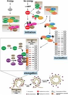
          Autophagy is a catabolic mechanism where intracellular material is degraded by vesicular structures called autophagolysosomes. Autophagy is necessary to maintain the normal function of the central nervous system (CNS), avoiding the accumulation of misfolded and aggregated proteins. Consistently, impaired autophagy has been associated with the pathogenesis of various neurodegenerative diseases. The proteins TAR DNA-binding protein-43 (TDP-43), which regulates RNA processing at different levels, and chromosome 9 open reading frame 72 (C9orf72), probably involved in membrane trafficking, are crucial in the development of neurodegenerative diseases such as Amyotrophic lateral sclerosis (ALS) and Frontotemporal Lobar Degeneration (FTLD). Additionally, recent studies have identified a role for these proteins in the control of autophagy. In this manuscript, we review what is known regarding the autophagic mechanism and discuss the involvement of TDP-43 and C9orf72 in autophagy and their impact on neurodegenerative diseases.

          Related collections

          Most cited references107

          • Record: found
          • Abstract: found
          • Article: found
          Is Open Access

          Autophagosome formation from membrane compartments enriched in phosphatidylinositol 3-phosphate and dynamically connected to the endoplasmic reticulum

          Autophagy is the engulfment of cytosol and organelles by double-membrane vesicles termed autophagosomes. Autophagosome formation is known to require phosphatidylinositol 3-phosphate (PI(3)P) and occurs near the endoplasmic reticulum (ER), but the exact mechanisms are unknown. We show that double FYVE domain–containing protein 1, a PI(3)P-binding protein with unusual localization on ER and Golgi membranes, translocates in response to amino acid starvation to a punctate compartment partially colocalized with autophagosomal proteins. Translocation is dependent on Vps34 and beclin function. Other PI(3)P-binding probes targeted to the ER show the same starvation-induced translocation that is dependent on PI(3)P formation and recognition. Live imaging experiments show that this punctate compartment forms near Vps34-containing vesicles, is in dynamic equilibrium with the ER, and provides a membrane platform for accumulation of autophagosomal proteins, expansion of autophagosomal membranes, and emergence of fully formed autophagosomes. This PI(3)P-enriched compartment may be involved in autophagosome biogenesis. Its dynamic relationship with the ER is consistent with the idea that the ER may provide important components for autophagosome formation.
            Bookmark
            • Record: found
            • Abstract: found
            • Article: not found

            Mitochondria supply membranes for autophagosome biogenesis during starvation.

            Starvation-induced autophagosomes engulf cytosol and/or organelles and deliver them to lysosomes for degradation, thereby resupplying depleted nutrients. Despite advances in understanding the molecular basis of this process, the membrane origin of autophagosomes remains unclear. Here, we demonstrate that, in starved cells, the outer membrane of mitochondria participates in autophagosome biogenesis. The early autophagosomal marker, Atg5, transiently localizes to punctae on mitochondria, followed by the late autophagosomal marker, LC3. The tail-anchor of an outer mitochondrial membrane protein also labels autophagosomes and is sufficient to deliver another outer mitochondrial membrane protein, Fis1, to autophagosomes. The fluorescent lipid NBD-PS (converted to NBD-phosphotidylethanolamine in mitochondria) transfers from mitochondria to autophagosomes. Photobleaching reveals membranes of mitochondria and autophagosomes are transiently shared. Disruption of mitochondria/ER connections by mitofusin2 depletion dramatically impairs starvation-induced autophagy. Mitochondria thus play a central role in starvation-induced autophagy, contributing membrane to autophagosomes. Copyright (c) 2010 Elsevier Inc. All rights reserved.
              Bookmark
              • Record: found
              • Abstract: found
              • Article: not found

              A subdomain of the endoplasmic reticulum forms a cradle for autophagosome formation.

              Autophagy is a bulk degradation process in eukaryotic cells and has fundamental roles in cellular homeostasis.The origin and source of autophagosomal membranes are long-standing questions in the field. Using electron microscopy, we show that, in mammalian culture cells, the endoplasmic reticulum (ER) associates with early autophagic structures called isolation membranes (IMs). Overexpression of an Atg4B mutant, which causes defects in autophagosome formation, induces the accumulation of ER-IM complexes. Electron tomography revealed that the ER-IM complex appears as a subdomain of the ER that formed a cradle encircling the IM, and showed that both ER and isolation membranes are interconnected.
                Bookmark

                Author and article information

                Contributors
                Journal
                Front Mol Neurosci
                Front Mol Neurosci
                Front. Mol. Neurosci.
                Frontiers in Molecular Neuroscience
                Frontiers Media S.A.
                1662-5099
                30 May 2017
                2017
                : 10
                : 170
                Affiliations
                [1] 1Dentistry Faculty, Institute in Dentistry Sciences, University of Chile Santiago, Chile
                [2] 2International Centre for Genetic Engineering and Biotechnology Trieste, Italy
                [3] 3Department of Physiology, Faculty of Biological Sciences, Pontificia Universidad Católica de Chile Santiago, Chile
                [4] 4Advanced Center for Chronic Diseases Santiago, Chile
                Author notes

                Edited by: Angelo Poletti, Università degli Studi di Milano, Italy

                Reviewed by: Alessandro Vercelli, University of Turin, Italy; Cristina Cereda, Fondazione Istituto Neurologico Nazionale Casimiro Mondino (IRCCS), Italy

                *Correspondence: Mauricio Budini mbudini@ 123456u.uchile.cl
                Article
                10.3389/fnmol.2017.00170
                5447761
                28611593
                3356e74b-8548-4be4-9ac3-6162a0c66833
                Copyright © 2017 Budini, Buratti, Morselli and Criollo.

                This is an open-access article distributed under the terms of the Creative Commons Attribution License (CC BY). The use, distribution or reproduction in other forums is permitted, provided the original author(s) or licensor are credited and that the original publication in this journal is cited, in accordance with accepted academic practice. No use, distribution or reproduction is permitted which does not comply with these terms.

                History
                : 01 February 2017
                : 15 May 2017
                Page count
                Figures: 7, Tables: 0, Equations: 0, References: 178, Pages: 21, Words: 15345
                Funding
                Funded by: Fondo Nacional de Desarrollo Científico y Tecnológico 10.13039/501100002850
                Award ID: 1161123
                Award ID: 1160820
                Award ID: 1140908
                Funded by: International Centre for Genetic Engineering and Biotechnology 10.13039/501100001688
                Award ID: CRP/CH113-04RG
                Award ID: CRP/CHL16-06
                Categories
                Neuroscience
                Review

                Neurosciences
                autophagy,neurodegenerative diseases,tdp-43,c9orf72,als,ftld
                Neurosciences
                autophagy, neurodegenerative diseases, tdp-43, c9orf72, als, ftld

                Comments

                Comment on this article