10
views
0
recommends
+1 Recommend
0 collections
    0
    shares
      • Record: found
      • Abstract: found
      • Article: found
      Is Open Access

      Novel RvD6 stereoisomer induces corneal nerve regeneration and wound healing post-injury by modulating trigeminal transcriptomic signature

      research-article

      Read this article at

      Bookmark
          There is no author summary for this article yet. Authors can add summaries to their articles on ScienceOpen to make them more accessible to a non-specialist audience.

          Abstract

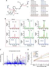
          The high-density corneal innervation plays a pivotal role in sustaining the integrity of the ocular surface. We have previously demonstrated that pigment epithelium-derived factor (PEDF) plus docosahexaenoic acid (DHA) promotes corneal nerve regeneration; here, we report the mechanism involved and the discovery of a stereospecific Resolvin D6-isomer (RvD6si) that drives the process. RvD6si promotes corneal wound healing and functional recovery by restoring corneal innervation after injury. RvD6si applied to the eye surface elicits a specific transcriptome signature in the trigeminal ganglion (TG) that includes Rictor, the rapamycin-insensitive complex-2 of mTOR (mTORC2), and genes involved in axon growth, whereas genes related to neuropathic pain are decreased. As a result, attenuation of ocular neuropathic pain and dry eye will take place. Thus, RvD6si opens up new therapeutic avenues for pathologies that affect corneal innervation.

          Related collections

          Most cited references41

          • Record: found
          • Abstract: found
          • Article: not found

          Corneal nerves: structure, contents and function

          Experimental Eye Research, 76(5), 521-542
            Bookmark
            • Record: found
            • Abstract: found
            • Article: found
            Is Open Access

            The role of calcitonin gene–related peptide in peripheral and central pain mechanisms including migraine

            Abstract Calcitonin gene–related peptide (CGRP) is a 37-amino acid peptide found primarily in the C and Aδ sensory fibers arising from the dorsal root and trigeminal ganglia, as well as the central nervous system. Calcitonin gene–related peptide was found to play important roles in cardiovascular, digestive, and sensory functions. Although the vasodilatory properties of CGRP are well documented, its somatosensory function regarding modulation of neuronal sensitization and of enhanced pain has received considerable attention recently. Growing evidence indicates that CGRP plays a key role in the development of peripheral sensitization and the associated enhanced pain. Calcitonin gene–related peptide is implicated in the development of neurogenic inflammation and it is upregulated in conditions of inflammatory and neuropathic pain. It is most likely that CGRP facilitates nociceptive transmission and contributes to the development and maintenance of a sensitized, hyperresponsive state not only of the primary afferent sensory neurons but also of the second-order pain transmission neurons within the central nervous system, thus contributing to central sensitization as well. The maintenance of a sensitized neuronal condition is believed to be an important factor underlying migraine. Recent successful clinical studies have shown that blocking the function of CGRP can alleviate migraine. However, the mechanisms through which CGRP may contribute to migraine are still not fully understood. We reviewed the role of CGRP in primary afferents, the dorsal root ganglion, and in the trigeminal system as well as its role in peripheral and central sensitization and its potential contribution to pain processing and to migraine.
              Bookmark
              • Record: found
              • Abstract: found
              • Article: not found

              Novel docosanoids inhibit brain ischemia-reperfusion-mediated leukocyte infiltration and pro-inflammatory gene expression.

              Ischemic stroke triggers lipid peroxidation and neuronal injury. Docosahexaenoic acid released from membrane phospholipids during brain ischemia is a major source of lipid peroxides. Leukocyte infiltration and pro-inflammatory gene expression also contribute to stroke damage. In this study using lipidomic analysis, we have identified stereospecific messengers from docosahexaenoate-oxygenation pathways in a mouse stroke model. Aspirin, widely used to prevent cerebrovascular disease, activates an additional pathway, which includes the 17R-resolvins. The newly discovered brain messenger 10,17S-docosatriene potently inhibited leukocyte infiltration, NFkappaB, and cyclooxygenase-2 induction in experimental stroke and elicited neuroprotection. In addition, in neural cells in culture, this lipid messenger also inhibited both interleukin 1-beta-induced NFkappaB activation and cyclooxygenase-2 expression. Thus, the specific novel bioactive docosanoids generated in vivo counteract leukocyte-mediated injury as well as pro-inflammatory gene induction. These results challenge the view that docosahexaenoate only participates in brain damage and demonstrate that this fatty acid is also the endogenous precursor to a neuroprotective signaling response to ischemia-reperfusion.
                Bookmark

                Author and article information

                Contributors
                hbazan1@lsuhsc.edu
                Journal
                Sci Rep
                Sci Rep
                Scientific Reports
                Nature Publishing Group UK (London )
                2045-2322
                12 March 2020
                12 March 2020
                2020
                : 10
                : 4582
                Affiliations
                ISNI 0000 0000 8954 1233, GRID grid.279863.1, Neuroscience Center of Excellence, School of Medicine, Louisiana State University Health New Orleans, ; New Orleans, LA 70112 USA
                Article
                61390
                10.1038/s41598-020-61390-8
                7067818
                32165657
                3345f135-c90a-4b2d-ae4b-6d17faf56e9f
                © The Author(s) 2020

                Open Access This article is licensed under a Creative Commons Attribution 4.0 International License, which permits use, sharing, adaptation, distribution and reproduction in any medium or format, as long as you give appropriate credit to the original author(s) and the source, provide a link to the Creative Commons license, and indicate if changes were made. The images or other third party material in this article are included in the article’s Creative Commons license, unless indicated otherwise in a credit line to the material. If material is not included in the article’s Creative Commons license and your intended use is not permitted by statutory regulation or exceeds the permitted use, you will need to obtain permission directly from the copyright holder. To view a copy of this license, visit http://creativecommons.org/licenses/by/4.0/.

                History
                : 13 December 2019
                : 24 February 2020
                Funding
                Funded by: FundRef http://dx.doi.org/10.13039/100005808, Vietnam Education Foundation;
                Funded by: FundRef http://dx.doi.org/10.13039/100000053, National Eye Institute;
                Award ID: R01EY19465
                Award Recipient :
                Categories
                Article
                Custom metadata
                © The Author(s) 2020

                Uncategorized
                molecular biology,neuroscience
                Uncategorized
                molecular biology, neuroscience

                Comments

                Comment on this article