1
views
0
recommends
+1 Recommend
0 collections
    0
    shares
      • Record: found
      • Abstract: found
      • Article: found
      Is Open Access

      A single genetic locus controls both expression of DPEP1/CHMP1A and kidney disease development via ferroptosis

      research-article

      Read this article at

      Bookmark
          There is no author summary for this article yet. Authors can add summaries to their articles on ScienceOpen to make them more accessible to a non-specialist audience.

          Abstract

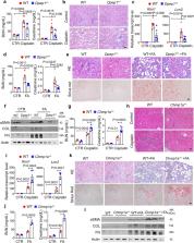
          Genome-wide association studies (GWAS) have identified loci for kidney disease, but the causal variants, genes, and pathways remain unknown. Here we identify two kidney disease genes Dipeptidase 1 ( DPEP1) and Charged Multivesicular Body Protein 1 A ( CHMP1A) via the triangulation of kidney function GWAS, human kidney expression, and methylation quantitative trait loci. Using single-cell chromatin accessibility and genome editing, we fine map the region that controls the expression of both genes. Mouse genetic models demonstrate the causal roles of both genes in kidney disease. Cellular studies indicate that both Dpep1 and Chmp1a are important regulators of a single pathway, ferroptosis and lead to kidney disease development via altering cellular iron trafficking.

          Abstract

          Identifying causal variants and genes is an essential step in interpreting GWAS loci. Here, the authors investigate a kidney disease GWAS locus with functional genomics data, CRISPR editing and mouse experiments to identify DPEP1 and CHMP1A as putative kidney disease genes via ferroptosis.

          Related collections

          Most cited references55

          • Record: found
          • Abstract: found
          • Article: not found

          Ferroptosis: an iron-dependent form of nonapoptotic cell death.

          Nonapoptotic forms of cell death may facilitate the selective elimination of some tumor cells or be activated in specific pathological states. The oncogenic RAS-selective lethal small molecule erastin triggers a unique iron-dependent form of nonapoptotic cell death that we term ferroptosis. Ferroptosis is dependent upon intracellular iron, but not other metals, and is morphologically, biochemically, and genetically distinct from apoptosis, necrosis, and autophagy. We identify the small molecule ferrostatin-1 as a potent inhibitor of ferroptosis in cancer cells and glutamate-induced cell death in organotypic rat brain slices, suggesting similarities between these two processes. Indeed, erastin, like glutamate, inhibits cystine uptake by the cystine/glutamate antiporter (system x(c)(-)), creating a void in the antioxidant defenses of the cell and ultimately leading to iron-dependent, oxidative death. Thus, activation of ferroptosis results in the nonapoptotic destruction of certain cancer cells, whereas inhibition of this process may protect organisms from neurodegeneration. Copyright © 2012 Elsevier Inc. All rights reserved.
            Bookmark
            • Record: found
            • Abstract: found
            • Article: not found

            Ferroptosis: A Regulated Cell Death Nexus Linking Metabolism, Redox Biology, and Disease

            Ferroptosis is a form of regulated cell death characterized by the iron-dependent accumulation of lipid hydroperoxides to lethal levels. Emerging evidence suggests that ferroptosis represents an ancient vulnerability caused by the incorporation of polyunsaturated fatty acids into cellular membranes, and cells have developed complex systems that exploit and defend against this vulnerability in different contexts. The sensitivity to ferroptosis is tightly linked to numerous biological processes, including amino acid, iron, and polyunsaturated fatty acid metabolism, and the biosynthesis of glutathione, phospholipids, NADPH, and coenzyme Q10. Ferroptosis has been implicated in the pathological cell death associated with degenerative diseases (i.e., Alzheimer's, Huntington's, and Parkinson's diseases), carcinogenesis, stroke, intracerebral hemorrhage, traumatic brain injury, ischemia-reperfusion injury, and kidney degeneration in mammals and is also implicated in heat stress in plants. Ferroptosis may also have a tumor-suppressor function that could be harnessed for cancer therapy. This Primer reviews the mechanisms underlying ferroptosis, highlights connections to other areas of biology and medicine, and recommends tools and guidelines for studying this emerging form of regulated cell death.
              Bookmark
              • Record: found
              • Abstract: found
              • Article: not found

              ACSL4 dictates ferroptosis sensitivity by shaping cellular lipid composition.

              Ferroptosis is a form of regulated necrotic cell death controlled by glutathione peroxidase 4 (GPX4). At present, mechanisms that could predict sensitivity and/or resistance and that may be exploited to modulate ferroptosis are needed. We applied two independent approaches-a genome-wide CRISPR-based genetic screen and microarray analysis of ferroptosis-resistant cell lines-to uncover acyl-CoA synthetase long-chain family member 4 (ACSL4) as an essential component for ferroptosis execution. Specifically, Gpx4-Acsl4 double-knockout cells showed marked resistance to ferroptosis. Mechanistically, ACSL4 enriched cellular membranes with long polyunsaturated ω6 fatty acids. Moreover, ACSL4 was preferentially expressed in a panel of basal-like breast cancer cell lines and predicted their sensitivity to ferroptosis. Pharmacological targeting of ACSL4 with thiazolidinediones, a class of antidiabetic compound, ameliorated tissue demise in a mouse model of ferroptosis, suggesting that ACSL4 inhibition is a viable therapeutic approach to preventing ferroptosis-related diseases.
                Bookmark

                Author and article information

                Contributors
                ksusztak@pennmedicine.upenn.edu
                Journal
                Nat Commun
                Nat Commun
                Nature Communications
                Nature Publishing Group UK (London )
                2041-1723
                23 August 2021
                23 August 2021
                2021
                : 12
                : 5078
                Affiliations
                [1 ]GRID grid.25879.31, ISNI 0000 0004 1936 8972, Department of Medicine, Renal Electrolyte and Hypertension Division, , Perelman School of Medicine, University of Pennsylvania, ; Philadelphia, PA 19104 USA
                [2 ]GRID grid.25879.31, ISNI 0000 0004 1936 8972, Department of Genetics, Perelman School of Medicine, , University of Pennsylvania, ; Philadelphia, PA 19104 USA
                [3 ]GRID grid.25879.31, ISNI 0000 0004 1936 8972, Graduate group in Genomics and Computational Biology, , Perelman School of Medicine, University of Pennsylvania, ; Philadelphia, PA 19104 USA
                [4 ]GRID grid.412282.f, ISNI 0000 0001 1091 2917, Division of Nephrology, Department of Internal Medicine, , University Hospital Carl Gustav Carus at the Technische Universität Dresden, ; Dresden, Germany
                [5 ]GRID grid.4488.0, ISNI 0000 0001 2111 7257, Biotechnology Center, Technische Universität Dresden, ; 01307 Dresden, Germany
                [6 ]Division of Genetic Medicine, Department of Medicine, Vanderbilt Genetics Institute, Nashville, TN 37232 USA
                [7 ]GRID grid.25879.31, ISNI 0000 0004 1936 8972, Department of Systems Pharmacology and Translational Therapeutics, , Perelman School of Medicine, University of Pennsylvania, ; Philadelphia, PA 19104 USA
                [8 ]GRID grid.25879.31, ISNI 0000 0004 1936 8972, Institute of Translational Medicine and Therapeutics, , Perelman School of Medicine, University of Pennsylvania, ; Philadelphia, PA 19104 USA
                Author information
                http://orcid.org/0000-0003-1866-270X
                http://orcid.org/0000-0002-1512-8993
                http://orcid.org/0000-0002-0733-8616
                http://orcid.org/0000-0002-3255-9517
                http://orcid.org/0000-0001-6287-9725
                http://orcid.org/0000-0001-7479-0920
                http://orcid.org/0000-0002-6205-9994
                http://orcid.org/0000-0002-1005-3726
                Article
                25377
                10.1038/s41467-021-25377-x
                8382756
                34426578
                32b64c78-ed20-46d2-bc06-e218db12968a
                © The Author(s) 2021

                Open Access This article is licensed under a Creative Commons Attribution 4.0 International License, which permits use, sharing, adaptation, distribution and reproduction in any medium or format, as long as you give appropriate credit to the original author(s) and the source, provide a link to the Creative Commons license, and indicate if changes were made. The images or other third party material in this article are included in the article’s Creative Commons license, unless indicated otherwise in a credit line to the material. If material is not included in the article’s Creative Commons license and your intended use is not permitted by statutory regulation or exceeds the permitted use, you will need to obtain permission directly from the copyright holder. To view a copy of this license, visit http://creativecommons.org/licenses/by/4.0/.

                History
                : 19 December 2020
                : 3 August 2021
                Funding
                Funded by: FundRef https://doi.org/10.13039/100000062, U.S. Department of Health & Human Services | NIH | National Institute of Diabetes and Digestive and Kidney Diseases (National Institute of Diabetes & Digestive & Kidney Diseases);
                Award ID: R01 DK076077
                Award ID: R01 DK087635
                Award Recipient :
                Funded by: U.S. Department of Health & Human Services | NIH | National Institute of Diabetes and Digestive and Kidney Diseases (National Institute of Diabetes & Digestive & Kidney Diseases)
                Categories
                Article
                Custom metadata
                © The Author(s) 2021

                Uncategorized
                genetics,functional genomics,kidney diseases
                Uncategorized
                genetics, functional genomics, kidney diseases

                Comments

                Comment on this article