25
views
0
recommends
+1 Recommend
0 collections
    0
    shares
      • Record: found
      • Abstract: found
      • Article: found
      Is Open Access

      Characterizing microglia activation: a spatial statistics approach to maximize information extraction

      research-article

      Read this article at

      Bookmark
          There is no author summary for this article yet. Authors can add summaries to their articles on ScienceOpen to make them more accessible to a non-specialist audience.

          Abstract

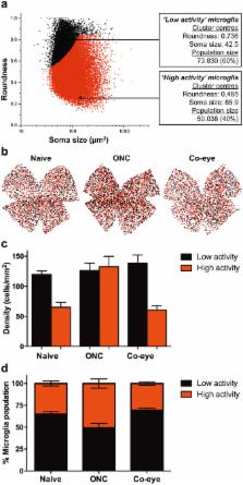
          Microglia play an important role in the pathology of CNS disorders, however, there remains significant uncertainty about the neuroprotective/degenerative role of these cells due to a lack of techniques to adequately assess their complex behaviour in response to injury. Advancing microscopy techniques, transgenic lines and well-characterized molecular markers, have made histological assessment of microglia populations more accessible. However, there is a distinct lack of tools to adequately extract information from these images to fully characterise microglia behaviour. This, combined with growing economic pressures and the ethical need to minimise the use of laboratory animals, led us to develop tools to maximise the amount of information obtained. This study describes a novel approach, combining image analysis with spatial statistical techniques. In addition to monitoring morphological parameters and global changes in microglia density, nearest neighbour distance, and regularity index, we used cluster analyses based on changes in soma size and roundness to yield novel insights into the behaviour of different microglia phenotypes in a murine optic nerve injury model. These methods should be considered a generic tool to quantitatively assess microglia activation, to profile phenotypic changes into microglia subpopulations, and to map spatial distributions in virtually every CNS region and disease state.

          Related collections

          Most cited references35

          • Record: found
          • Abstract: found
          • Article: not found

          Microglia development and function.

          Proper development and function of the mammalian central nervous system (CNS) depend critically on the activity of parenchymal sentinels referred to as microglia. Although microglia were first described as ramified brain-resident phagocytes, research conducted over the past century has expanded considerably upon this narrow view and ascribed many functions to these dynamic CNS inhabitants. Microglia are now considered among the most versatile cells in the body, possessing the capacity to morphologically and functionally adapt to their ever-changing surroundings. Even in a resting state, the processes of microglia are highly dynamic and perpetually scan the CNS. Microglia are in fact vital participants in CNS homeostasis, and dysregulation of these sentinels can give rise to neurological disease. In this review, we discuss the exciting developments in our understanding of microglial biology, from their developmental origin to their participation in CNS homeostasis and pathophysiological states such as neuropsychiatric disorders, neurodegeneration, sterile injury responses, and infectious diseases. We also delve into the world of microglial dynamics recently uncovered using real-time imaging techniques.
            Bookmark
            • Record: found
            • Abstract: found
            • Article: not found

            Endocytosis by random initiation and stabilization of clathrin-coated pits.

            Clathrin-coated vesicles carry traffic from the plasma membrane to endosomes. We report here the real-time visualization of cargo sorting and endocytosis by clathrin-coated pits in living cells. We have detected the formation of coats by monitoring incorporation of fluorescently tagged clathrin or its adaptor AP-2; we have also followed clathrin-mediated uptake of transferrin and of single LDL or reovirus particles. The intensity of a cargo-loaded clathrin cluster grows steadily during its lifetime, and the time required to complete assembly is proportional to the size of the cargo particle. These results are consistent with a nucleation-growth mechanism and an approximately constant growth rate. There are no strongly preferred nucleation sites. A proportion of the nucleation events are weak and short lived. Cargo incorporation occurs primarily or exclusively in a newly formed coated pit. Our data lead to a model in which coated pits initiate randomly but collapse unless stabilized, perhaps by cargo capture.
              Bookmark
              • Record: found
              • Abstract: found
              • Article: found
              Is Open Access

              Quantitating the subtleties of microglial morphology with fractal analysis

              It is well established that microglial form and function are inextricably linked. In recent years, the traditional view that microglial form ranges between “ramified resting” and “activated amoeboid” has been emphasized through advancing imaging techniques that point to microglial form being highly dynamic even within the currently accepted morphological categories. Moreover, microglia adopt meaningful intermediate forms between categories, with considerable crossover in function and varying morphologies as they cycle, migrate, wave, phagocytose, and extend and retract fine and gross processes. From a quantitative perspective, it is problematic to measure such variability using traditional methods, but one way of quantitating such detail is through fractal analysis. The techniques of fractal analysis have been used for quantitating microglial morphology, to categorize gross differences but also to differentiate subtle differences (e.g., amongst ramified cells). Multifractal analysis in particular is one technique of fractal analysis that may be useful for identifying intermediate forms. Here we review current trends and methods of fractal analysis, focusing on box counting analysis, including lacunarity and multifractal analysis, as applied to microglial morphology.
                Bookmark

                Author and article information

                Contributors
                lies.degroef@kuleuven.be
                Journal
                Sci Rep
                Sci Rep
                Scientific Reports
                Nature Publishing Group UK (London )
                2045-2322
                8 May 2017
                8 May 2017
                2017
                : 7
                : 1576
                Affiliations
                [1 ]ISNI 0000000121901201, GRID grid.83440.3b, Glaucoma and Retinal Neurodegenerative Disease Research Group, Institute of Ophthalmology, , University College London, ; 11-43 Bath Street, London, EC1V 9EL United Kingdom
                [2 ]ISNI 0000 0001 0668 7884, GRID grid.5596.f, Neural Circuit Development and Regeneration Research Group, Department of Biology, , University of Leuven, ; Naamsestraat 61 box 2464, 3000 Leuven, Belgium
                [3 ]ISNI 0000 0001 0693 2181, GRID grid.417895.6, Western Eye Hospital, , Imperial College Healthcare NHS Trust, ; 171 Marylebone Road, London, NW1 5QH United Kingdom
                Author information
                http://orcid.org/0000-0003-0186-1411
                http://orcid.org/0000-0002-3329-3474
                Article
                1747
                10.1038/s41598-017-01747-8
                5431479
                28484229
                3195bcf4-8bfe-4e7d-a3e9-d7ec3fbe0e53
                © The Author(s) 2017

                Open Access This article is licensed under a Creative Commons Attribution 4.0 International License, which permits use, sharing, adaptation, distribution and reproduction in any medium or format, as long as you give appropriate credit to the original author(s) and the source, provide a link to the Creative Commons license, and indicate if changes were made. The images or other third party material in this article are included in the article’s Creative Commons license, unless indicated otherwise in a credit line to the material. If material is not included in the article’s Creative Commons license and your intended use is not permitted by statutory regulation or exceeds the permitted use, you will need to obtain permission directly from the copyright holder. To view a copy of this license, visit http://creativecommons.org/licenses/by/4.0/.

                History
                : 19 January 2017
                : 30 March 2017
                Categories
                Article
                Custom metadata
                © The Author(s) 2017

                Uncategorized
                Uncategorized

                Comments

                Comment on this article