191
views
0
recommends
+1 Recommend
0 collections
    0
    shares
      • Record: found
      • Abstract: found
      • Article: found
      Is Open Access

      The Sedoheptulose Kinase CARKL Directs Macrophage Polarization through Control of Glucose Metabolism

      research-article

      Read this article at

      Bookmark
          There is no author summary for this article yet. Authors can add summaries to their articles on ScienceOpen to make them more accessible to a non-specialist audience.

          Summary

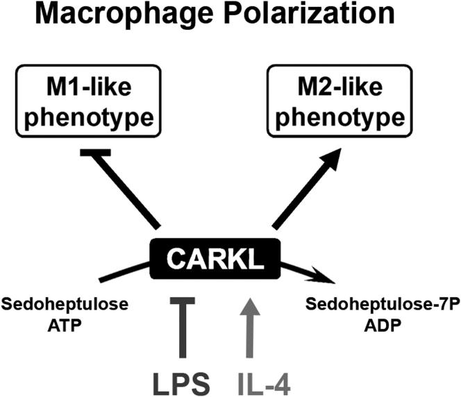
          Immune cells are somewhat unique in that activation responses can alter quantitative phenotypes upwards of 100,000-fold. To date little is known about the metabolic adaptations necessary to mount such dramatic phenotypic shifts. Screening for novel regulators of macrophage activation, we found nonprotein kinases of glucose metabolism among the most enriched classes of candidate immune modulators. We find that one of these, the carbohydrate kinase- like protein CARKL, is rapidly downregulated in vitro and in vivo upon LPS stimulation in both mice and humans. Interestingly, CARKL catalyzes an orphan reaction in the pentose phosphate pathway, refocusing cellular metabolism to a high-redox state upon physiological or artificial downregulation. We find that CARKL-dependent metabolic reprogramming is required for proper M1- and M2-like macrophage polarization and uncover a rate-limiting requirement for appropriate glucose flux in macrophage polarization.

          Abstract

          Graphical Abstract

          Highlights

          ► Screened 199 human kinases for their immunoregulatory potential ► CARKL bridges glycolysis, the pentose phosphate pathway, and immune function ► CARKL focuses cellular metabolism toward a “high-redox” state ► CARKL regulation is required for macrophage polarization

          Related collections

          Most cited references36

          • Record: found
          • Abstract: not found
          • Article: not found

          On the origin of cancer cells.

          O WARBURG (1956)
            Bookmark
            • Record: found
            • Abstract: found
            • Article: not found

            The inflammasomes: guardians of the body.

            The innate immune system relies on its capacity to rapidly detect invading pathogenic microbes as foreign and to eliminate them. The discovery of Toll-like receptors (TLRs) provided a class of membrane receptors that sense extracellular microbes and trigger antipathogen signaling cascades. More recently, intracellular microbial sensors have been identified, including NOD-like receptors (NLRs). Some of the NLRs also sense nonmicrobial danger signals and form large cytoplasmic complexes called inflammasomes that link the sensing of microbial products and metabolic stress to the proteolytic activation of the proinflammatory cytokines IL-1beta and IL-18. The NALP3 inflammasome has been associated with several autoinflammatory conditions including gout. Likewise, the NALP3 inflammasome is a crucial element in the adjuvant effect of aluminum and can direct a humoral adaptive immune response. In this review, we discuss the role of NLRs, and in particular the inflammasomes, in the recognition of microbial and danger components and the role they play in health and disease.
              Bookmark
              • Record: found
              • Abstract: found
              • Article: found

              p53 regulates glucose metabolism through an IKK-NF-kappaB pathway and inhibits cell transformation.

              Cancer cells use aerobic glycolysis preferentially for energy provision and this metabolic change is important for tumour growth. Here, we have found a link between the tumour suppressor p53, the transcription factor NF-kappaB and glycolysis. In p53-deficient primary cultured cells, kinase activities of IKKalpha and IKKbeta and subsequent NF-kappaB activity were enhanced. Activation of NF-kappaB, by loss of p53, caused an increase in the rate of aerobic glycolysis and upregulation of Glut3. Oncogenic Ras-induced cell transformation and acceleration of aerobic glycolysis in p53-deficient cells were suppressed in the absence of p65/NF-kappaB expression, and were restored by GLUT3 expression. It was also shown that a glycolytic inhibitor diminished the enhanced IKK activity in p53-deficient cells. Moreover, in Ras-expressing p53-deficient cells, IKK activity was suppressed by p65 deficiency and restored by GLUT3 expression. Taken together, these data indicate that p53 restricts activation of the IKK-NF-kappaB pathway through suppression of glycolysis. These results suggest that a positive-feedback loop exists, whereby glycolysis drives IKK-NF-kappaB activation, and that hyperactivation of this loop by loss of p53 is important in oncogene-induced cell transformation.
                Bookmark

                Author and article information

                Journal
                Cell Metab
                Cell Metab
                Cell Metabolism
                Cell Press
                1550-4131
                1932-7420
                06 June 2012
                06 June 2012
                : 15
                : 6
                : 813-826
                Affiliations
                [1 ]Department of Laboratory Medicine, Medical University of Vienna, A-1090, Austria
                [2 ]Department of Pathophysiology and Allergy Research, Medical University of Vienna, A-1090, Austria
                [3 ]Center for Medical Statistics, Informatics and Intelligent Systems, Department for Biosimulation and Bioinformatics, Medical University of Vienna, A-1090, Austria
                [4 ]Center for Physiology and Pharmacology, Institute for Vascular Biology and Thrombosis Research, Medical University of Vienna, A-1090, Austria
                [5 ]Department of Pediatrics, Medical University of Vienna, A-1090, Austria
                [6 ]Harvard Medical School and the Transplant Institute, Department of Surgery, Beth Israel Deaconess Medical Center, Boston, MA 05215, USA
                [7 ]Department of Chemistry, University of Natural Resources and Applied Life Sciences, Vienna, A-1190, Austria
                [8 ]Institute of Pharmacology and Toxicology, Department of Biomedical Sciences, University of Veterinary Medicine Vienna, Vienna, A-1210, Austria
                [9 ]Department of Internal Medicine, University of Michigan, Ann Arbor, MI 48109, USA
                [10 ]Harvard Institute of Proteomics, Harvard Medical School, Cambridge, MA 02115, USA
                [11 ]Max Planck Institute of Immunobiology and Epigenetics, 79108 Freiburg, Germany
                Author notes
                []Corresponding author arvand.haschemi@ 123456meduniwien.ac.at
                [∗∗ ]Corresponding author pospisilik@ 123456immunbio.mpg.de
                [12]

                These authors contributed equally to this work

                [13]

                Present address: Agilent Technologies, Santa Clara, CA 95051, USA

                [14]

                Present address: Janssen Research & Development, Radnor, PA 19087, USA

                Article
                CMET1125
                10.1016/j.cmet.2012.04.023
                3370649
                22682222
                3141badd-b92a-4058-a656-0b6c751f8b3e
                © 2012 ELL & Excerpta Medica.

                This document may be redistributed and reused, subject to certain conditions.

                History
                : 21 June 2011
                : 18 November 2011
                : 26 April 2012
                Categories
                Article

                Cell biology
                Cell biology

                Comments

                Comment on this article