13
views
0
recommends
+1 Recommend
0 collections
    0
    shares
      • Record: found
      • Abstract: found
      • Article: found
      Is Open Access

      Berberine suppresses apoptosis and extracellular matrix (ECM) degradation in nucleus pulposus cells and ameliorates disc degeneration in a rodent model

      research-article

      Read this article at

      Bookmark
          There is no author summary for this article yet. Authors can add summaries to their articles on ScienceOpen to make them more accessible to a non-specialist audience.

          Abstract

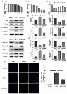
          Intervertebral disc degeneration (IVDD) is a chronic disease with complicated pathology involving nucleus pulposus (NP) cell apoptosis and extracellular matrix (ECM) degradation. Previous studies have shown that moderate autophagy has a protective effect against apoptosis in NP cells. Berberine (BBR) is an alkaloid compound with many beneficial properties including antimicrobial, anti-inflammatory, antioxidative, and anti-apoptotic activity. Recently, it was found to induce autophagy in various tissues as well. Thus, we hypothesized that BBR may exert a therapeutic effect on IVDD through autophagy activation. In this study, we investigated the effects of BBR on IVDD and delineated a potential mechanism. BBR treatment in vitro inhibited the expression of pro-apoptotic proteins induced by tert-butyl hydroperoxide (TBHP), and increased the expression of anti-apoptotic Bcl-2. Furthermore, it prevented ECM degradation by inhibiting the production of matrix-degrading enzymes. Additionally, BBR treatment significantly activated autophagy in NP cells. However, autophagy inhibition markedly suppressed BBR's effects on NP cell apoptosis and ECM degeneration, indicating that autophagy activation with BBR treatment is protective against IVDD. In vivo, BBR treatment increased the expression of LC3 in disc cells and prevented the development of IVDD in a needle puncture-induced rat model. Thus, BBR stimulates autophagy as a protective mechanism against NP cell apoptosis and ECM degeneration, revealing its therapeutic potential in the treatment of IVDD.

          Related collections

          Most cited references25

          • Record: found
          • Abstract: found
          • Article: not found

          Autophagy is a protective mechanism in normal cartilage, and its aging-related loss is linked with cell death and osteoarthritis.

          Autophagy is a process for turnover of intracellular organelles and molecules that protects cells during stress responses. We undertook this study to evaluate the potential roles of Unc-51-like kinase 1 (ULK1), an inducer of autophagy, Beclin1, a regulator of autophagy, and microtubule-associated protein 1 light chain 3 (LC3), which executes autophagy, in the development of osteoarthritis (OA) and in cartilage cell death. Expression of ULK1, Beclin1, and LC3 was analyzed in normal and OA human articular cartilage and in knee joints of mice with aging-related and surgically induced OA, using immunohistochemistry and Western blotting. Poly(ADP-ribose) polymerase (PARP) p85 expression was used to determine the correlation between cell death and autophagy. ULK1, Beclin1, and LC3 were constitutively expressed in normal human articular cartilage. ULK1, Beclin1, and LC3 protein expression was reduced in OA chondrocytes and cartilage, but these 3 proteins were strongly expressed in the OA cell clusters. In mouse knee joints, loss of glycosaminoglycans (GAGs) was observed at ages 9 months and 12 months and in the surgical OA model, 8 weeks after knee destabilization. Expression of ULK1, Beclin1, and LC3 decreased together with GAG loss, while PARP p85 expression was increased. Autophagy may be a protective or homeostatic mechanism in normal cartilage. In contrast, human OA and aging-related and surgically induced OA in mice are associated with a reduction and loss of ULK1, Beclin1, and LC3 expression and a related increase in apoptosis. These results suggest that compromised autophagy represents a novel mechanism in the development of OA.
            Bookmark
            • Record: found
            • Abstract: found
            • Article: found
            Is Open Access

            ROS: Crucial Intermediators in the Pathogenesis of Intervertebral Disc Degeneration

            Excessive reactive oxygen species (ROS) generation in degenerative intervertebral disc (IVD) indicates the contribution of oxidative stress to IVD degeneration (IDD), giving a novel insight into the pathogenesis of IDD. ROS are crucial intermediators in the signaling network of disc cells. They regulate the matrix metabolism, proinflammatory phenotype, apoptosis, autophagy, and senescence of disc cells. Oxidative stress not only reinforces matrix degradation and inflammation, but also promotes the decrease in the number of viable and functional cells in the microenvironment of IVDs. Moreover, ROS modify matrix proteins in IVDs to cause oxidative damage of disc extracellular matrix, impairing the mechanical function of IVDs. Consequently, the progression of IDD is accelerated. Therefore, a therapeutic strategy targeting oxidative stress would provide a novel perspective for IDD treatment. Various antioxidants have been proposed as effective drugs for IDD treatment. Antioxidant supplementation suppresses ROS production in disc cells to promote the matrix synthesis of disc cells and to prevent disc cells from death and senescence in vitro. However, there is not enough in vivo evidence to support the efficiency of antioxidant supplementation to retard the process of IDD. Further investigations based on in vivo and clinical studies will be required to develop effective antioxidative therapies for IDD.
              Bookmark
              • Record: found
              • Abstract: found
              • Article: not found

              Cell death in intervertebral disc degeneration.

              Degeneration of intervertebral disc (IVD) is mainly a chronic process of excessive destruction of the extracellular matrix (ECM), and also is thought to be the primary cause of low back pain. Presently, however, the underlying mechanism of IVD degeneration is still not elucidated. Cellular loss from cell death has been believed to contribute to the degradation of ECM and plays an important role in the process of IVD degeneration, but the mechanisms of cell death in degenerated IVD remain unclear. Apoptosis, a very important type of IVD cell death, has been considered to play a crucial role in the process of degeneration. Autophagy, a non-apoptosis death type of programmed cell death, has been considered extensively involved in many pathological and physiological processes, including the degenerative diseases. Thus, the research on cell death in IVD degeneration has become a new focus recently. In this review, by analyzing the available literature pertaining to cell death in IVD and discussing the inducing factors of IVD degeneration, NP cells and ECM in IVD degeneration, apoptotic signal transduction pathways involved in IVD cell death, the relationship of cell death with IVD degeneration and potential therapeutic strategy for IVD degeneration by regulating cell death, we conclude that different stimuli induce cell death in IVD via various signal transduction pathways, and that cell death may play a key role in the degenerative process of IVD. Regulation of cell death could be a potential and attractive therapeutic strategy for IVD degeneration.
                Bookmark

                Author and article information

                Journal
                Int J Biol Sci
                Int. J. Biol. Sci
                ijbs
                International Journal of Biological Sciences
                Ivyspring International Publisher (Sydney )
                1449-2288
                2018
                30 April 2018
                : 14
                : 6
                : 682-692
                Affiliations
                [1 ]Department of Orthopaedic Surgery, The Second Affiliated Hospital and Yuying Children's Hospital of Wenzhou Medical University, 325027 Wenzhou, China
                [2 ]Molecular Pharmacology Research Center, School of Pharmaceutical Sciences, Wenzhou Medical University, 325035, Wenzhou, China
                [3 ]Department of Molecular Pharmacology, Albert Einstein College of Medicine, Bronx, NY 10461, U.S.A.
                [4 ]Department of Orthopaedic Surgery, Ruian Peoples Hospital & the 3rd Hospital Affiliated to Wenzhou Medical University, 325200 Ruian, China
                Author notes
                ✉ Corresponding authors: Jian Xiao, Molecular Pharmacology Research Center, School of Pharmaceutical Sciences, Wenzhou Medical University, 325035, Wenzhou, China. E-mail: xfxj2000@ 123456126.com and Zhouguang Wang, Molecular Pharmacology Research Center, School of Pharmaceutical Sciences, Wenzhou Medical University, 325035, Wenzhou, China. Tel.:+86-577-85773087; E-mail: wzhouguang@ 123456gmail.com and Xiangyang Wang, Department of Orthopaedic Surgery, The Second Affiliated Hospital of Wenzhou Medical University, Xueyuan Xi Road, Wenzhou, Zhejiang, 325027, China. Tel.: 86-577-88829799; Email: xiangyangwang@ 123456126.com

                # These authors contributed equally to this work.

                Competing Interests: The authors have declared that no competing interest exists.

                Article
                ijbsv14p0682
                10.7150/ijbs.24081
                6001656
                29904282
                2fe6c6e5-1c28-4010-aecf-2f909c852fed
                © Ivyspring International Publisher

                This is an open access article distributed under the terms of the Creative Commons Attribution (CC BY-NC) license ( https://creativecommons.org/licenses/by-nc/4.0/). See http://ivyspring.com/terms for full terms and conditions.

                History
                : 29 November 2017
                : 13 February 2018
                Categories
                Research Paper

                Life sciences
                Life sciences

                Comments

                Comment on this article