5
views
0
recommends
+1 Recommend
0 collections
    0
    shares
      • Record: found
      • Abstract: found
      • Article: found
      Is Open Access

      High resolution cryo-EM structure of the helical RNA-bound Hantaan virus nucleocapsid reveals its assembly mechanisms

      research-article

      Read this article at

      Bookmark
          There is no author summary for this article yet. Authors can add summaries to their articles on ScienceOpen to make them more accessible to a non-specialist audience.

          Abstract

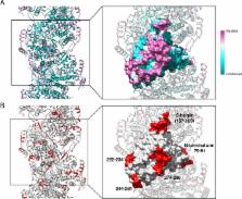
          Negative-strand RNA viruses condense their genome into helical nucleocapsids that constitute essential templates for viral replication and transcription. The intrinsic flexibility of nucleocapsids usually prevents their full-length structural characterisation at high resolution. Here, we describe purification of full-length recombinant metastable helical nucleocapsid of Hantaan virus ( Hantaviridae family, Bunyavirales order) and determine its structure at 3.3 Å resolution by cryo-electron microscopy. The structure reveals the mechanisms of helical multimerisation via sub-domain exchanges between protomers and highlights nucleotide positions in a continuous positively charged groove compatible with viral genome binding. It uncovers key sites for future structure-based design of antivirals that are currently lacking to counteract life-threatening hantavirus infections. The structure also suggests a model of nucleoprotein-polymerase interaction that would enable replication and transcription solely upon local disruption of the nucleocapsid.

          eLife digest

          Rats and mice sometimes transmit hantaviruses, a family of microbes that can cause deadly human diseases. For example, the Hantaan virus leads to haemorrhagic fevers that are potentially fatal. There are no vaccine or even drugs against these infections.

          To multiply, viruses must insert their genetic material inside a cell. While the body often detects and destroys foreign genetic information, hantaviruses can still evade our defences. Molecules called nucleoproteins bind to the viral genome, hiding it away in long helices called nucleocapsids. When the virus needs to replicate, an enzyme opens up the nucleocapsid, reads and copies the genetic code, and then closes the helix. Yet, researchers know little about the details of this process, or even the structure of the nucleocapsid.

          Here, Arragain et al. use a method called cryo-electron microscopy to examine and piece together the exact 3D structure of the Hantaan virus nucleocapsid. This was possible because the new technique allows scientists to observe biological molecules at an unprecedented, near atomic resolution. The resulting model reveals that the viral genome nests into a groove inside the nucleocapsid. It also shows that specific interactions between nucleoproteins stabilise the helix. Finally, the model helps to provide hypotheses on how the enzyme could read the genome without breaking the capsid.

          Mapping out the structure and the interactions of the nucleocapsid is the first step towards finding molecules that could destabilise the helix and neutralise the virus: this could help fight both the Hantaan virus and other members of its deadly family.

          Related collections

          Most cited references24

          • Record: found
          • Abstract: found
          • Article: not found

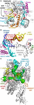
          Organization of the influenza virus replication machinery.

          Influenza virus ribonucleoprotein complexes (RNPs) are central to the viral life cycle and in adaptation to new host species. RNPs are composed of the viral genome, viral polymerase, and many copies of the viral nucleoprotein. In vitro cell expression of all RNP protein components with four of the eight influenza virus gene segments enabled structural determination of native influenza virus RNPs by means of cryogenic electron microscopy (cryo-EM). The cryo-EM structure reveals the architecture and organization of the native RNP, defining the attributes of its largely helical structure and how polymerase interacts with nucleoprotein and the viral genome. Observations of branched-RNP structures in negative-stain electron microscopy and their putative identification as replication intermediates suggest a mechanism for viral replication by a second polymerase on the RNP template.
            Bookmark
            • Record: found
            • Abstract: found
            • Article: found
            Is Open Access

            Structural Insights into Bunyavirus Replication and Its Regulation by the vRNA Promoter

            Summary Segmented negative-strand RNA virus (sNSV) polymerases transcribe and replicate the viral RNA (vRNA) within a ribonucleoprotein particle (RNP). We present cryo-EM and X-ray structures of, respectively, apo- and vRNA bound La Crosse orthobunyavirus (LACV) polymerase that give atomic-resolution insight into how such RNPs perform RNA synthesis. The complementary 3′ and 5′ vRNA extremities are sequence specifically bound in separate sites on the polymerase. The 5′ end binds as a stem-loop, allosterically structuring functionally important polymerase active site loops. Identification of distinct template and product exit tunnels allows proposal of a detailed model for template-directed replication with minimal disruption to the circularised RNP. The similar overall architecture and vRNA binding of monomeric LACV to heterotrimeric influenza polymerase, despite high sequence divergence, suggests that all sNSV polymerases have a common evolutionary origin and mechanism of RNA synthesis. These results will aid development of replication inhibitors of diverse, serious human pathogenic viruses.
              Bookmark
              • Record: found
              • Abstract: found
              • Article: not found

              Structure and assembly of the Ebola virus nucleocapsid

              Ebola and Marburg viruses are filoviruses: filamentous, enveloped viruses that cause hemorrhagic fever1. Filoviruses are within the order Mononegavirales2 which also includes rabies virus, measles virus, and respiratory syncytial virus. Mononegaviruses have non-segmented, single-stranded negative-sense RNA genomes that are encapsidated by nucleoprotein (NP) and other viral proteins to form a helical nucleocapsid (NC). NC acts as a scaffold for virus assembly and as a template for genome transcription and replication. Insights into NP-NP interactions have been derived from structural studies of oligomerized, RNA-encapsidating NP3–6 and cryo-electron microscopy (cryo-EM) of NC7–12 or NC-like structures11–13. There have been no high-resolution reconstructions of complete mononegavirus NCs. Here, we have applied cryo-electron tomography and subtomogram averaging to determine the structure of Ebola virus NC within intact viruses and recombinant NC-like assemblies. These structures reveal the identity and arrangement of the NC components, and suggest that the formation of an extended alpha-helix from the disordered C-terminal region of NP-core links NP oligomerization, NC condensation, RNA encapsidation, and accessory protein recruitment.
                Bookmark

                Author and article information

                Contributors
                Role: Reviewing Editor
                Role: Senior Editor
                Journal
                eLife
                Elife
                eLife
                eLife
                eLife Sciences Publications, Ltd
                2050-084X
                14 January 2019
                2019
                : 8
                : e43075
                Affiliations
                [1 ]deptElectron Microscopy and Methods Group Université Grenoble Alpes, CNRS, CEA, Institute for Structural Biology GrenobleFrance
                [2 ]deptComplexes Macromoléculaires Viraux Aix-Marseille Université, CNRS, INSERM, AFMB UMR 7257 MarseilleFrance
                University of Virginia United States
                University of California, Berkeley United States
                University of Virginia United States
                Author information
                http://orcid.org/0000-0002-5593-4682
                http://orcid.org/0000-0003-4977-7948
                http://orcid.org/0000-0002-6525-5042
                http://orcid.org/0000-0002-1908-3921
                http://orcid.org/0000-0002-1459-3201
                https://orcid.org/0000-0002-2834-7386
                Article
                43075
                10.7554/eLife.43075
                6365055
                30638449
                2fcb5825-5ff9-40a4-94ad-f02003df0783
                © 2019, Arragain et al

                This article is distributed under the terms of the Creative Commons Attribution License, which permits unrestricted use and redistribution provided that the original author and source are credited.

                History
                : 23 October 2018
                : 10 January 2019
                Funding
                Funded by: FundRef http://dx.doi.org/10.13039/100012952, Université Grenoble Alpes;
                Award ID: G7H-IRS17H50
                Award Recipient :
                The funders had no role in study design, data collection and interpretation, or the decision to submit the work for publication.
                Categories
                Short Report
                Structural Biology and Molecular Biophysics
                Custom metadata
                Hantaan virus nucleocapsid cryo-EM structure determined at 3.3 Å resolution reveals how nucleoproteins assemble into a metastable helix containing a continuous RNA-binding groove compatible with genome encapsidation and compaction.

                Life sciences
                bunyavirales,hantavirus,hantaan,nucleocapsid,nucleoprotein,replication,virus
                Life sciences
                bunyavirales, hantavirus, hantaan, nucleocapsid, nucleoprotein, replication, virus

                Comments

                Comment on this article