11
views
0
recommends
+1 Recommend
0 collections
    0
    shares
      • Record: found
      • Abstract: found
      • Article: found
      Is Open Access

      Cardiac troponin T associates with left ventricular function and synchrony assessed by CMR in the general population: results from the Akershus Cardiac Examination 1950 Study

      research-article

      Read this article at

      Bookmark
          There is no author summary for this article yet. Authors can add summaries to their articles on ScienceOpen to make them more accessible to a non-specialist audience.

          Abstract

          Background and aim

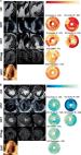
          Cardiac troponin T (cTnT) is a blood biomarker of myocardial injury that is associated with future adverse cardiovascular events in the general population. Left ventricular (LV) global longitudinal strain (GLS) and mechanical dispersion (MD) are metrics of systolic function and synchrony that can be obtained from cardiac imaging. Studies suggest an association between cTnT and echocardiographically assessed GLS and MD, but it is unknown whether cTnT relates to these metrics when assessed by cardiac magnetic resonance (CMR). We hypothesized that cTnT associates with GLS and with MD assessed by CMR feature tracking (CMR-FT) in the general population.

          Methods and results

          cTnT and CMR-FT measurements were performed in 186 community dwellers from the Akershus Cardiac Examination 1950 Study. The participants’ age ranged from 68 to 70 years. Median cTnT concentration was 7.0 ng/L (interquartile interval 5.0–12.6 ng/L), median absolute value of GLS was 17.3% (interquartile interval 15.7–18.8%), and median MD was 80.7 milliseconds (interquartile interval 61.8–105.0 milliseconds). In multivariable linear regression models adjusted for common clinical risk factors of cardiovascular disease, with GLS and MD as outcome and cTnT as the predictor variable of interest, log 10 transformed cTnT was significantly associated with both absolute GLS [β-coefficient −1.65, confidence interval (−2.84, −0.46)] and MD [β-coefficient 28.56, confidence interval (12.14, 44.92)].

          Conclusion

          In older adults from the general population, higher cTnT concentrations are associated with worse systolic function and synchrony assessed by CMR-FT LV GLS and MD, adding information about myocardial function to traditional risk factors.

          Graphical Abstract

          Graphical Abstract

          In older adults from the general population, higher cardiac troponin T is associated with worse global longitudinal strain and worse mechanical dispersion as assessed by cardiac magnetic resonance feature tracking.

          Related collections

          Most cited references27

          • Record: found
          • Abstract: not found
          • Article: not found

          Fourth Universal Definition of Myocardial Infarction (2018).

            Bookmark
            • Record: found
            • Abstract: found
            • Article: found
            Is Open Access

            Myocardial strain imaging: review of general principles, validation, and sources of discrepancies

            Abstract Myocardial tissue tracking imaging techniques have been developed for a more accurate evaluation of myocardial deformation (i.e. strain), with the potential to overcome the limitations of ejection fraction (EF) and to contribute, incremental to EF, to the diagnosis and prognosis in cardiac diseases. While most of the deformation imaging techniques are based on the similar principles of detecting and tracking specific patterns within an image, there are intra- and inter-imaging modality inconsistencies limiting the wide clinical applicability of strain. In this review, we aimed to describe the particularities of the echocardiographic and cardiac magnetic resonance deformation techniques, in order to understand the discrepancies in strain measurement, focusing on the potential sources of variation: related to the software used to analyse the data, to the different physics of image acquisition and the different principles of 2D vs. 3D approaches. As strain measurements are not interchangeable, it is highly desirable to work with validated strain assessment tools, in order to derive information from evidence-based data. There is, however, a lack of solid validation of the current tissue tracking techniques, as only a few of the commercial deformation imaging softwares have been properly investigated. We have, therefore, addressed in this review the neglected issue of suboptimal validation of tissue tracking techniques, in order to advocate for this matter.
              Bookmark
              • Record: found
              • Abstract: found
              • Article: found
              Is Open Access

              Principles of cardiovascular magnetic resonance feature tracking and echocardiographic speckle tracking for informed clinical use

              Tissue tracking technology of routinely acquired cardiovascular magnetic resonance (CMR) cine acquisitions has increased the apparent ease and availability of non-invasive assessments of myocardial deformation in clinical research and practice. Its widespread availability thanks to the fact that this technology can in principle be applied on images that are part of every CMR or echocardiographic protocol. However, the two modalities are based on very different methods of image acquisition and reconstruction, each with their respective strengths and limitations. The image tracking methods applied are not necessarily directly comparable between the modalities, or with those based on dedicated CMR acquisitions for strain measurement such as tagging or displacement encoding. Here we describe the principles underlying the image tracking methods for CMR and echocardiography, and the translation of the resulting tracking estimates into parameters suited to describe myocardial mechanics. Technical limitations are presented with the objective of suggesting potential solutions that may allow informed and appropriate use in clinical applications.
                Bookmark

                Author and article information

                Contributors
                Journal
                Eur Heart J Imaging Methods Pract
                Eur Heart J Imaging Methods Pract
                ehjimp
                European Heart Journal. Imaging Methods and Practice
                Oxford University Press (UK )
                2755-9637
                July 2024
                30 July 2024
                30 July 2024
                : 2
                : 3
                : qyae078
                Affiliations
                K.G. Jebsen Center for Cardiac Biomarkers, Institute of Clinical Medicine, University of Oslo , Campus Akershus University Hospital, P.b. 1000 NO-1478 Lørenskog, Norway
                Department of Diagnostic Imaging, Akershus University Hospital , Sykehusveien 25, Nordbyhagen, 1478 Lørenskog, Norway
                Department of Diagnostic Imaging, Akershus University Hospital , Sykehusveien 25, Nordbyhagen, 1478 Lørenskog, Norway
                K.G. Jebsen Center for Cardiac Biomarkers, Institute of Clinical Medicine, University of Oslo , Campus Akershus University Hospital, P.b. 1000 NO-1478 Lørenskog, Norway
                Department of Medical Research, Vestre Viken Bærum Hospital , Gjettum, Norway
                Department of Medical Research, Vestre Viken Bærum Hospital , Gjettum, Norway
                Institute of Clinical Medicine, University of Oslo , Oslo, Norway
                K.G. Jebsen Center for Cardiac Biomarkers, Institute of Clinical Medicine, University of Oslo , Campus Akershus University Hospital, P.b. 1000 NO-1478 Lørenskog, Norway
                Division for Research and Innovation, Akershus Clinical Research Center, Akershus University Hospital , Lørenskog, Norway
                K.G. Jebsen Center for Cardiac Biomarkers, Institute of Clinical Medicine, University of Oslo , Campus Akershus University Hospital, P.b. 1000 NO-1478 Lørenskog, Norway
                Department of Infectious Diseases, Division of Medicine, Akershus University Hospital , Lørenskog, Norway
                K.G. Jebsen Center for Cardiac Biomarkers, Institute of Clinical Medicine, University of Oslo , Campus Akershus University Hospital, P.b. 1000 NO-1478 Lørenskog, Norway
                Department of Cardiology, Akershus University Hospital , Lørenskog, Norway
                K.G. Jebsen Center for Cardiac Biomarkers, Institute of Clinical Medicine, University of Oslo , Campus Akershus University Hospital, P.b. 1000 NO-1478 Lørenskog, Norway
                Department of Diagnostic Imaging, Akershus University Hospital , Sykehusveien 25, Nordbyhagen, 1478 Lørenskog, Norway
                Author notes
                Corresponding author. E-mail: s.l.heck@ 123456medisin.uio.no

                Conflict of interest: The authors have no conflicts of interest related to this work. Unrelated to this work, T.O. has received funding from Abbott Diagnostics; nonfinancial support from Novartis, Abbott Diagnostics, Roche Diagnostics, and SomaLogic; and honoraria from Siemens Healthineers, Roche Diagnostics, and Abbott Diagnostics. Unrelated to this work, J.S. serves as medical advisor for SiFi labs (unpaid).

                Author information
                https://orcid.org/0000-0001-7813-3143
                https://orcid.org/0009-0002-0404-1777
                https://orcid.org/0000-0002-7139-5252
                https://orcid.org/0000-0002-7681-8621
                https://orcid.org/0000-0002-7223-5138
                https://orcid.org/0000-0003-2114-3134
                https://orcid.org/0000-0002-5994-9304
                https://orcid.org/0000-0002-6452-0369
                https://orcid.org/0000-0002-5430-1613
                Article
                qyae078
                10.1093/ehjimp/qyae078
                11441316
                39351316
                2f89442b-273b-4b7f-bd2d-69e663a9fc1b
                © The Author(s) 2024. Published by Oxford University Press on behalf of the European Society of Cardiology.

                This is an Open Access article distributed under the terms of the Creative Commons Attribution-NonCommercial License ( https://creativecommons.org/licenses/by-nc/4.0/), which permits non-commercial re-use, distribution, and reproduction in any medium, provided the original work is properly cited. For commercial re-use, please contact reprints@oup.com for reprints and translation rights for reprints. All other permissions can be obtained through our RightsLink service via the Permissions link on the article page on our site—for further information please contact journals.permissions@oup.com.

                History
                : 25 April 2024
                : 25 July 2024
                : 20 August 2024
                Page count
                Pages: 9
                Funding
                Funded by: Akershus University Hospital, DOI 10.13039/501100012446;
                Funded by: Norwegian Health Association, DOI 10.13039/501100013263;
                Funded by: Norwegian Research Council, DOI 10.13039/501100005416;
                Funded by: Roche, DOI 10.13039/100004337;
                Categories
                Original Article
                AcademicSubjects/MED00010
                AcademicSubjects/MED00160
                AcademicSubjects/MED00200
                AcademicSubjects/MED00870
                Eurheartj/31
                Eurheartj/33
                Eurheartj/45
                Eurheartj/47

                cardiac troponin t,global longitudinal strain,mechanical dispersion,lv systolic function,lv synchrony,cardiac magnetic resonance feature tracking

                Comments

                Comment on this article

                scite_
                0
                0
                0
                0
                Smart Citations
                0
                0
                0
                0
                Citing PublicationsSupportingMentioningContrasting
                View Citations

                See how this article has been cited at scite.ai

                scite shows how a scientific paper has been cited by providing the context of the citation, a classification describing whether it supports, mentions, or contrasts the cited claim, and a label indicating in which section the citation was made.

                Similar content121

                Most referenced authors862