0
views
0
recommends
+1 Recommend
0 collections
    0
    shares
      • Record: found
      • Abstract: found
      • Article: found
      Is Open Access

      Changes of in vivo electrical conductivity in the brain and torso related to age, fat fraction and sex using MRI

      research-article

      Read this article at

      Bookmark
          There is no author summary for this article yet. Authors can add summaries to their articles on ScienceOpen to make them more accessible to a non-specialist audience.

          Abstract

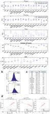
          This work was inspired by the observation that a majority of MR-electrical properties tomography studies are based on direct comparisons with ex vivo measurements carried out on post-mortem samples in the 90’s. As a result, the in vivo conductivity values obtained from MRI in the megahertz range in different types of tissues (brain, liver, tumors, muscles, etc.) found in the literature may not correspond to their ex vivo equivalent, which still serves as a reference for electromagnetic modelling. This study aims to pave the way for improving current databases since the definition of personalized electromagnetic models (e.g. for Specific Absorption Rate estimation) would benefit from better estimation. Seventeen healthy volunteers underwent MRI of both brain and thorax/abdomen using a three-dimensional ultrashort echo-time (UTE) sequence. We estimated conductivity (S/m) in several classes of macroscopic tissue using a customized reconstruction method from complex UTE images, and give general statistics for each of these regions (mean-median-standard deviation). These values are used to find possible correlations with biological parameters such as age, sex, body mass index and/or fat volume fraction, using linear regression analysis. In short, the collected in vivo values show significant deviations from the ex vivo values in conventional databases, and we show significant relationships with the latter parameters in certain organs for the first time, e.g. a decrease in brain conductivity with age.

          Related collections

          Most cited references75

          • Record: found
          • Abstract: found
          • Article: not found

          nnU-Net: a self-configuring method for deep learning-based biomedical image segmentation

          Biomedical imaging is a driver of scientific discovery and a core component of medical care and is being stimulated by the field of deep learning. While semantic segmentation algorithms enable image analysis and quantification in many applications, the design of respective specialized solutions is non-trivial and highly dependent on dataset properties and hardware conditions. We developed nnU-Net, a deep learning-based segmentation method that automatically configures itself, including preprocessing, network architecture, training and post-processing for any new task. The key design choices in this process are modeled as a set of fixed parameters, interdependent rules and empirical decisions. Without manual intervention, nnU-Net surpasses most existing approaches, including highly specialized solutions on 23 public datasets used in international biomedical segmentation competitions. We make nnU-Net publicly available as an out-of-the-box tool, rendering state-of-the-art segmentation accessible to a broad audience by requiring neither expert knowledge nor computing resources beyond standard network training.
            Bookmark
            • Record: found
            • Abstract: found
            • Article: not found

            The dielectric properties of biological tissues: II. Measurements in the frequency range 10 Hz to 20 GHz.

            Three experimental techniques based on automatic swept-frequency network and impedance analysers were used to measure the dielectric properties of tissue in the frequency range 10 Hz to 20 GHz. The technique used in conjunction with the impedance analyser is described. Results are given for a number of human and animal tissues, at body temperature, across the frequency range, demonstrating that good agreement was achieved between measurements using the three pieces of equipment. Moreover, the measured values fall well within the body of corresponding literature data.
              Bookmark
              • Record: found
              • Abstract: found
              • Article: not found

              Adaptive reconstruction of phased array MR imagery

              An adaptive implementation of the spatial matched filter and its application to the reconstruction of phased array MR imagery is described. Locally relevant array correlation statistics for the NMR signal and noise processes are derived directly from the set of complex individual coil images, in the form of sample correlation matrices. Eigen-analysis yields an optimal filter vector for the estimated signal and noise array correlation statistics. The technique enables near-optimal reconstruction of multicoil MR imagery without a-priori knowledge of the individual coil field maps or noise correlation structure. Experimental results indicate SNR performance approaching that of the optimal matched filter. Compared to the sum-of-squares technique, the RMS noise level in dark image regions is reduced by as much as the square root of N, where N is the number of coils in the array. The technique is also effective in suppressing localized motion and flow artifacts. Copyright 2000 Wiley-Liss, Inc.
                Bookmark

                Author and article information

                Contributors
                paul.soullie@univ-lorraine.fr
                Journal
                Sci Rep
                Sci Rep
                Scientific Reports
                Nature Publishing Group UK (London )
                2045-2322
                12 July 2024
                12 July 2024
                2024
                : 14
                : 16109
                Affiliations
                [1 ]GRID grid.29172.3f, ISNI 0000 0001 2194 6418, IADI U1254, , INSERM and Université de Lorraine, ; Nancy, France
                [2 ]GRID grid.426119.9, ISNI 0000 0004 0621 9441, Siemens Healthcare SAS, ; Saint Denis, France
                [3 ]CIC-IT 1433, INSERM, Université de Lorraine and CHRU Nancy, ( https://ror.org/02vjkv261) Nancy, France
                Article
                67014
                10.1038/s41598-024-67014-9
                11245625
                38997324
                2f2ffef0-7c1c-4ee1-b11a-100611d18c46
                © The Author(s) 2024

                Open Access This article is licensed under a Creative Commons Attribution 4.0 International License, which permits use, sharing, adaptation, distribution and reproduction in any medium or format, as long as you give appropriate credit to the original author(s) and the source, provide a link to the Creative Commons licence, and indicate if changes were made. The images or other third party material in this article are included in the article's Creative Commons licence, unless indicated otherwise in a credit line to the material. If material is not included in the article's Creative Commons licence and your intended use is not permitted by statutory regulation or exceeds the permitted use, you will need to obtain permission directly from the copyright holder. To view a copy of this licence, visit http://creativecommons.org/licenses/by/4.0/.

                History
                : 15 April 2024
                : 8 July 2024
                Categories
                Article
                Custom metadata
                © Springer Nature Limited 2024

                Uncategorized
                predictive markers,magnetic resonance imaging,biological physics
                Uncategorized
                predictive markers, magnetic resonance imaging, biological physics

                Comments

                Comment on this article