0
views
0
recommends
+1 Recommend
0 collections
    0
    shares
      • Record: found
      • Abstract: found
      • Article: found
      Is Open Access

      Surgical management of syringomyelia associated with spinal arachnoid web: strategies and outcomes

      research-article

      Read this article at

      Bookmark
          There is no author summary for this article yet. Authors can add summaries to their articles on ScienceOpen to make them more accessible to a non-specialist audience.

          Abstract

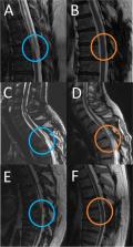
          Spinal arachnoid web (SAW) is a rare disease entity characterized as band-like arachnoid tissue that can cause spinal cord compression and syringomyelia. This study aimed to analyze the surgical management of the spinal arachnoid web in patients with syringomyelia, focusing on surgical strategies and outcomes. A total of 135 patients with syringomyelia underwent surgery at our department between November 2003 and December 2022. All patients underwent magnetic resonance imaging (MRI), with a special syringomyelia protocol (including TrueFISP and CINE), and electrophysiology. Among these patients, we searched for patients with SAW with syringomyelia following careful analysis of neuroradiological data and surgical reports. The criteria for SAW were as follows: displacement of the spinal cord, disturbed but preserved CSF flow, and intraoperative arachnoid web. Patients were evaluated for initial symptoms, surgical strategies, and complications by reviewing surgical reports, patient documents, neuroradiological data, and follow-up data. Of the 135 patients, 3 (2.22%) fulfilled the SAW criteria. The mean patient age was 51.67 ± 8.33 years. Two patients were male, and one was female. The affected levels were T2/3, T6, and T8. Excision of the arachnoid web was performed in all cases. No significant change in intraoperative monitoring was noted. Postoperatively, none of the patients presented new neurological symptoms. The MRI 3 months after surgery revealed that the syringomyelia improved in all cases, and caliber variation of the spinal cord could not be detected anymore. All clinical symptoms improved. In summary, SAW can be safely treated by surgery. Even though syringomyelia usually improves on MRI and symptoms also improve, residual symptoms might be observed. We advocate for clear criteria for the diagnosis of SAW and a standardized diagnostic (MRI including TrueFISP and CINE).

          Related collections

          Most cited references31

          • Record: found
          • Abstract: found
          • Article: not found

          Unraveling the riddle of syringomyelia.

          Dan Greitz (2006)
          The pathophysiology of syringomyelia development is not fully understood. Current prevailing theories suggest that increased pulse pressure in the subarachnoid space forces cerebrospinal fluid (CSF) through the spinal cord into the syrinx. It is generally accepted that the syrinx consists of CSF. The here-proposed intramedullary pulse pressure theory instead suggests that syringomyelia is caused by increased pulse pressure in the spinal cord and that the syrinx consists of extracellular fluid. A new principle is introduced implying that the distending force in the production of syringomyelia is a relative increase in pulse pressure in the spinal cord compared to that in the nearby subarachnoid space. The formation of a syrinx then occurs by the accumulation of extracellular fluid in the distended cord. A previously unrecognized mechanism for syrinx formation, the Bernoulli theorem, is also described. The Bernoulli theorem or the Venturi effect states that the regional increase in fluid velocity in a narrowed flow channel decreases fluid pressure. In Chiari I malformations, the systolic CSF pulse pressure and downward motion of the cerebellar tonsils are significantly increased. This leads to increased spinal CSF velocities and, as a consequence of the Bernoulli theorem, decreased fluid pressure in narrow regions of the spinal CSF pathways. The resulting relatively low CSF pressure in the narrowed CSF pathway causes a suction effect on the spinal cord that distends the cord during each systole. Syringomyelia develops by the accumulation of extracellular fluid in the distended cord. In posttraumatic syringomyelia, the downwards directed systolic CSF pulse pressure is transmitted and reflected into the spinal cord below and above the traumatic subarachnoid blockage, respectively. The ensuing increase in intramedullary pulse pressure distends the spinal cord and causes syringomyelia on both sides of the blockage. The here-proposed concept has the potential to unravel the riddle of syringomyelia and affords explanations to previously unanswered clinical and theoretical problems with syringomyelia. It also explains why syringomyelia associated with Chiari I malformations may develop in any part of the spinal cord including the medullary conus. Syringomyelia thus preferentially develops where the systolic CSF flow causes a suction effect on the spinal cord, i.e., at or immediately caudal to physiological or pathological encroachments of the spinal subarachnoid space.
            Bookmark
            • Record: found
            • Abstract: found
            • Article: not found

            The fine anatomy of the human spinal meninges. A light and scanning electron microscopy study.

            The fine anatomy of the human spinal meninges was examined in five postmortem spinal cords taken within 12 hours after death from patients aged 15 months to 46 years. Specimens of spinal cord were viewed in transverse section and from the dorsal and ventral aspects by scanning electron microscopy. Transverse sections of spinal cord and meninges were also examined by light microscopy. The arachnoid mater was seen to be closely applied to the inner aspect of the dura. An intermediate fenestrated leptomeningeal layer was observed attached to the inner aspect of the arachnoid mater and was reflected ventrally to form a series of dorsal septa. As it arborized laterally over the surface of the cord to surround nerves and blood vessels, the intermediate layer became highly fenestrated but remained distinct from the pia and arachnoid mater. The pia mater appeared to form a continuous layer which was reflected off the surface of the cord to coat blood vessels within the subarachnoid space in a manner similar to that described in the leptomeninges over the human cerebral cortex. Each dentate ligament consisted of a collagenous core which was continuous with the subpial connective tissue and was attached at intervals to the dura; pia-arachnoid cells coated the surface of the dentate ligaments. The present study suggests that the fine anatomy of the human spinal meninges differs significantly from that described in other mammals.
              Bookmark
              • Record: found
              • Abstract: found
              • Article: not found

              Pathophysiology of primary spinal syringomyelia.

              The pathogenesis of syringomyelia in patients with an associated spinal lesion is incompletely understood. The authors hypothesized that in primary spinal syringomyelia, a subarachnoid block effectively shortens the length of the spinal subarachnoid space (SAS), reducing compliance and the ability of the spinal theca to dampen the subarachnoid CSF pressure waves produced by brain expansion during cardiac systole. This creates exaggerated spinal subarachnoid pressure waves during every heartbeat that act on the spinal cord above the block to drive CSF into the spinal cord and create a syrinx. After a syrinx is formed, enlarged subarachnoid pressure waves compress the external surface of the spinal cord, propel the syrinx fluid, and promote syrinx progression.
                Bookmark

                Author and article information

                Contributors
                sasan_adib2002@yahoo.de
                jens.schittenhelm@med.uni-tuebingen.de
                p.kurucz@klinikum-stuttgart.de
                till-karsten.hauser@med.uni-tuebingen.de
                marcos.tatagiba@med.uni-tuebingen.de
                Journal
                Neurosurg Rev
                Neurosurg Rev
                Neurosurgical Review
                Springer Berlin Heidelberg (Berlin/Heidelberg )
                0344-5607
                1437-2320
                26 June 2023
                26 June 2023
                2023
                : 46
                : 1
                : 152
                Affiliations
                [1 ]GRID grid.10392.39, ISNI 0000 0001 2190 1447, Department of Neurosurgery, , University of Tuebingen, ; Hoppe-Seyler-Straße 3, 72076 Tuebingen, Germany
                [2 ]GRID grid.10392.39, ISNI 0000 0001 2190 1447, Department of Neuropathology, , University of Tuebingen, ; Hoppe-Seyler-Straße 3, 72076 Tuebingen, Germany
                [3 ]GRID grid.419842.2, ISNI 0000 0001 0341 9964, Department of Neurosurgery, , Klinikum Stuttgart, ; Kriegsbergstraße 60, 70174 Stuttgart, Germany
                [4 ]GRID grid.10392.39, ISNI 0000 0001 2190 1447, Department of Neuroradiology, , University of Tuebingen, ; Hoppe-Seyler-Straße 3, 72076 Tuebingen, Germany
                Author information
                https://orcid.org/0000-0002-1879-8490
                Article
                2071
                10.1007/s10143-023-02071-8
                10293323
                37358703
                2ef6707c-e2dd-4da7-be3e-a56b0e06ec7f
                © The Author(s) 2023

                Open Access This article is licensed under a Creative Commons Attribution 4.0 International License, which permits use, sharing, adaptation, distribution and reproduction in any medium or format, as long as you give appropriate credit to the original author(s) and the source, provide a link to the Creative Commons licence, and indicate if changes were made. The images or other third party material in this article are included in the article's Creative Commons licence, unless indicated otherwise in a credit line to the material. If material is not included in the article's Creative Commons licence and your intended use is not permitted by statutory regulation or exceeds the permitted use, you will need to obtain permission directly from the copyright holder. To view a copy of this licence, visit http://creativecommons.org/licenses/by/4.0/.

                History
                : 26 February 2023
                : 22 May 2023
                : 20 June 2023
                Funding
                Funded by: Universitätsklinikum Tübingen (8868)
                Categories
                Research
                Custom metadata
                © Springer-Verlag GmbH Germany, part of Springer Nature 2023

                Surgery
                spinal arachnoid web,syringomyelia,scalpel sign,csf flow,venturi effect,arachnopathy
                Surgery
                spinal arachnoid web, syringomyelia, scalpel sign, csf flow, venturi effect, arachnopathy

                Comments

                Comment on this article