7
views
0
recommends
+1 Recommend
0 collections
    0
    shares
      • Record: found
      • Abstract: found
      • Article: found
      Is Open Access

      Class V chitin synthase and β(1,3)-glucan synthase co-travel in the same vesicle in Zymoseptoria tritici

      other

      Read this article at

      Bookmark
          There is no author summary for this article yet. Authors can add summaries to their articles on ScienceOpen to make them more accessible to a non-specialist audience.

          Highlights

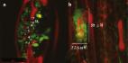
          • Native chitin (Chs5) and glucan synthase (Gsc1) visualised in the pathogen Zymoseptoria tritici.

          • Chs5 and Gsc1 are transported along microtubules.

          • Chs5 and Gsc1 do localise to the apical plasma membrane, but not the Spitzenkörper.

          • Light and electron microscopy how co-travel of Chs5 and Gsc1 in the same secretory vesicle.

          • Enzyme delivery in Z. tritici is different from Neurospora crassa, but similar to Ustilago maydis.

          Abstract

          The fungal cell wall consists of proteins and polysaccharides, formed by the co-ordinated activity of enzymes, such as chitin or glucan synthases. These enzymes are delivered via secretory vesicles to the hyphal tip. In the ascomycete Neurospora crassa, chitin synthases and β(1,3)-glucan synthase are transported in different vesicles, whereas they co-travel along microtubules in the basidiomycete Ustilago maydis. This suggests fundamental differences in wall synthesis between taxa. Here, we visualize the class V chitin synthase ZtChs5 and the β(1,3)-glucan synthase ZtGcs1 in the ascomycete Zymoseptoria tritici. Live cell imaging demonstrate that both enzymes co-locate to the apical plasma membrane, but are not concentrated in the Spitzenkörper. Delivery involves co-transport along microtubules of the chitin and glucan synthase. Live cell imaging and electron microscopy suggest that both cell wall synthases locate in the same vesicle. Thus, microtubule-dependent co-delivery of cell wall synthases in the same vesicle is found in asco- and basidiomycetes.

          Related collections

          Most cited references37

          • Record: found
          • Abstract: not found
          • Article: not found

          The fungi.

            Bookmark
            • Record: found
            • Abstract: found
            • Article: not found

            Confocal microscopy of FM4-64 as a tool for analysing endocytosis and vesicle trafficking in living fungal hyphae.

            Confocal microscopy of amphiphilic styryl dyes has been used to investigate endocytosis and vesicle trafficking in living fungal hyphae. Hyphae were treated with FM4-64, FM1-43 or TMA-DPH, three of the most commonly used membrane-selective dyes reported as markers of endocytosis. All three dyes were rapidly internalized within hyphae. FM4-64 was found best for imaging the dynamic changes in size, morphology and position of the apical vesicle cluster within growing hyphal tips because of its staining pattern, greater photostability and low cytotoxicity. FM4-64 was taken up into both the apical and subapical compartments of living hyphae in a time-dependent manner. The pattern of stain distribution was broadly similar in a range of fungal species tested (Aspergillus nidulans, Botrytis cinerea, Magnaporthe grisea, Neurospora crassa, Phycomyces blakesleeanus, Puccinia graminis, Rhizoctonia solani, Sclerotinia sclerotiorum and Trichoderma viride). With time, FM4-64 was internalized from the plasma membrane appearing in structures corresponding to putative endosomes, the apical vesicle cluster, the vacuolar membrane and mitochondria. These observations are consistent with dye internalization by endocytosis. A speculative model of the vesicle trafficking network within growing hyphae is presented.
              Bookmark
              • Record: found
              • Abstract: found
              • Article: found
              Is Open Access

              The impact of Septoria tritici Blotch disease on wheat: An EU perspective

              Highlights • Zymospetoria tritici is a threat to wheat production in the EU Z.t.’s plastic genome increases the potential severity of this threat in the future. • Climate change may also affect the risk from Z.t. • We estimate the spore numbers produced by Z.t. during each infection cycle. • We calculate 1) the economic value of wheat in the three main EU producers 2) the cost of and economic return for fungicide treatment of wheat vs Z.t.
                Bookmark

                Author and article information

                Contributors
                Journal
                Fungal Genet Biol
                Fungal Genet Biol
                Fungal Genetics and Biology
                Academic Press
                1087-1845
                1096-0937
                1 February 2020
                February 2020
                : 135
                : 103286
                Affiliations
                [a ]School of Biosciences, University of Exeter, Exeter EX4 4QD, UK
                [b ]65 Vallcalent Street, 25006 Lleida, Spain
                Author notes
                [* ]Corresponding author. G.Steinberg@ 123456exeter.ac.uk
                [1]

                These author contributed equally.

                Article
                S1087-1845(19)30289-0 103286
                10.1016/j.fgb.2019.103286
                7967022
                31672687
                2dedb96b-bd3d-4571-9840-539553614eb8
                © 2019 The Authors

                This is an open access article under the CC BY license (http://creativecommons.org/licenses/by/4.0/).

                History
                : 16 September 2019
                : 15 October 2019
                : 17 October 2019
                Categories
                Video Article

                Plant science & Botany
                membrane trafficking,chitin synthases,glucan synthase,cell wall formation

                Comments

                Comment on this article