26
views
0
recommends
+1 Recommend
0 collections
    0
    shares
      • Record: found
      • Abstract: found
      • Article: found
      Is Open Access

      Regulation of Placental Extravillous Trophoblasts by the Maternal Uterine Environment

      review-article

      Read this article at

      Bookmark
          There is no author summary for this article yet. Authors can add summaries to their articles on ScienceOpen to make them more accessible to a non-specialist audience.

          Abstract

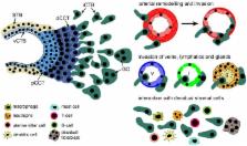
          During placentation invasive extravillous trophoblasts (EVTs) migrate into the maternal uterus and modify its vessels. In particular, remodeling of the spiral arteries by EVTs is critical for adapting blood flow and nutrient transport to the developing fetus. Failures in this process have been noticed in different pregnancy complications such as preeclampsia, intrauterine growth restriction, stillbirth, or recurrent abortion. Upon invasion into the decidua, the endometrium of pregnancy, EVTs encounter different maternal cell types such as decidual macrophages, uterine NK (uNK) cells and stromal cells expressing a plethora of growth factors and cytokines. Here, we will summarize development of the EVT lineage, a process occurring independently of the uterine environment, and formation of its different subtypes. Further, we will discuss interactions of EVTs with arteries, veins and lymphatics and illustrate how the decidua and its different immune cells regulate EVT differentiation, invasion and survival. The present literature suggests that the decidual environment and its soluble factors critically modulate EVT function and reproductive success.

          Related collections

          Most cited references237

          • Record: found
          • Abstract: found
          • Article: not found

          Tissue-Resident Macrophage Ontogeny and Homeostasis.

          Defining the origins and developmental pathways of tissue-resident macrophages should help refine our understanding of the role of these cells in various disease settings and enable the design of novel macrophage-targeted therapies. In recent years the long-held belief that macrophage populations in the adult are continuously replenished by monocytes from the bone marrow (BM) has been overturned with the advent of new techniques to dissect cellular ontogeny. The new paradigm suggests that several tissue-resident macrophage populations are seeded during waves of embryonic hematopoiesis and self-maintain independently of BM contribution during adulthood. However, the exact nature of the embryonic progenitors that give rise to adult tissue-resident macrophages is still debated, and the mechanisms enabling macrophage population maintenance in the adult are undefined. Here, we review the emergence of these concepts and discuss current controversies and future directions in macrophage biology.
            Bookmark
            • Record: found
            • Abstract: found
            • Article: not found

            Local macrophage proliferation, rather than recruitment from the blood, is a signature of TH2 inflammation.

            A defining feature of inflammation is the accumulation of innate immune cells in the tissue that are thought to be recruited from the blood. We reveal that a distinct process exists in which tissue macrophages undergo rapid in situ proliferation in order to increase population density. This inflammatory mechanism occurred during T helper 2 (T(H)2)-related pathologies under the control of the archetypal T(H)2 cytokine interleukin-4 (IL-4) and was a fundamental component of T(H)2 inflammation because exogenous IL-4 was sufficient to drive accumulation of tissue macrophages through self-renewal. Thus, expansion of innate cells necessary for pathogen control or wound repair can occur without recruitment of potentially tissue-destructive inflammatory cells.
              Bookmark
              • Record: found
              • Abstract: found
              • Article: not found

              Modulation of macrophage phenotype by cell shape.

              Phenotypic polarization of macrophages is regulated by a milieu of cues in the local tissue microenvironment. Although much is known about how soluble factors influence macrophage polarization, relatively little is known about how physical cues present in the extracellular environment might modulate proinflammatory (M1) vs. prohealing (M2) activation. Specifically, the role of cell shape has not been explored, even though it has been observed that macrophages adopt different geometries in vivo. We and others observed that macrophages polarized toward different phenotypes in vitro exhibit dramatic changes in cell shape: M2 cells exhibit an elongated shape compared with M1 cells. Using a micropatterning approach to control macrophage cell shape directly, we demonstrate here that elongation itself, without exogenous cytokines, leads to the expression of M2 phenotype markers and reduces the secretion of inflammatory cytokines. Moreover, elongation enhances the effects of M2-inducing cytokines IL-4 and IL-13 and protects cells from M1-inducing stimuli LPS and IFN-γ. In addition shape- but not cytokine-induced polarization is abrogated when actin and actin/myosin contractility are inhibited by pharmacological agents, suggesting a role for the cytoskeleton in the control of macrophage polarization by cell geometry. Our studies demonstrate that alterations in cell shape associated with changes in ECM architecture may provide integral cues to modulate macrophage phenotype polarization.
                Bookmark

                Author and article information

                Contributors
                Journal
                Front Immunol
                Front Immunol
                Front. Immunol.
                Frontiers in Immunology
                Frontiers Media S.A.
                1664-3224
                13 November 2018
                2018
                : 9
                : 2597
                Affiliations
                [1] 1Department of Obstetrics and Gynaecology, Medical University of Vienna , Vienna, Austria
                [2] 2British Columbia's Children's Hospital Research Institute , Vancouver, BC, Canada
                [3] 3Department of Obstetrics and Gynecology, University of British Columbia , Vancouver, BC, Canada
                Author notes

                Edited by: Nandor Gabor Than, Hungarian Academy of Sciences (MTA), Hungary

                Reviewed by: Caroline Dunk, Lunenfeld-Tanenbaum Research Institute, Canada; Joanna James, University of Auckland, New Zealand

                *Correspondence: Martin Knöfler martin.knoefler@ 123456meduniwien.ac.at

                This article was submitted to Immunological Tolerance and Regulation, a section of the journal Frontiers in Immunology

                Article
                10.3389/fimmu.2018.02597
                6243063
                30483261
                2d98c0f6-b5ad-4a77-8953-b4e5122e7902
                Copyright © 2018 Pollheimer, Vondra, Baltayeva, Beristain and Knöfler.

                This is an open-access article distributed under the terms of the Creative Commons Attribution License (CC BY). The use, distribution or reproduction in other forums is permitted, provided the original author(s) and the copyright owner(s) are credited and that the original publication in this journal is cited, in accordance with accepted academic practice. No use, distribution or reproduction is permitted which does not comply with these terms.

                History
                : 28 August 2018
                : 22 October 2018
                Page count
                Figures: 3, Tables: 0, Equations: 0, References: 272, Pages: 18, Words: 16859
                Funding
                Funded by: Austrian Science Fund 10.13039/501100002428
                Funded by: Oesterreichische Nationalbank 10.13039/501100004061
                Funded by: Canadian Institutes of Health Research 10.13039/501100000024
                Categories
                Immunology
                Review

                Immunology
                placental development,extravillous trophoblast,decidual immune cells,trophoblast invasion,uterine natural killer cells,decidual macrophages

                Comments

                Comment on this article