22
views
0
recommends
+1 Recommend
0 collections
    0
    shares
      • Record: found
      • Abstract: found
      • Article: found
      Is Open Access

      Fungal Malignant Otitis Externa Involves a Cascade of Complications Culminating in Pseudoaneurysm of Internal Maxillary Artery: A Case Report

      research-article

      Read this article at

      Bookmark
          There is no author summary for this article yet. Authors can add summaries to their articles on ScienceOpen to make them more accessible to a non-specialist audience.

          Abstract

          Patient: Male, 66

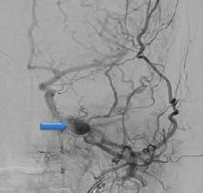
          Final Diagnosis: Pseudoaneurysm of internal maxillary artery secondary to fungal malignant otitis externa

          Symptoms: Epistaxis

          Medication: —

          Clinical Procedure: Functional endoscopic sinus surgery, endovascular embolization

          Specialty: Infectious Diseases

          Objective:

          Unusual clinical course

          Background:

          Pseudomonal infection is the most common cause of malignant otitis externa (MOE), which typically affects elderly diabetic patients. Fungi are a rare cause of MOE. MOE can be life-threatening if not recognized and treated promptly. It can result in a wide spectrum of complications, including skull-base osteomyelitis, cranial nerve palsy, cerebral venous thrombosis, and brain abscess. Pseudoaneurysm formation of the intracranial vessels is a life-threatening complication of MOE that is seldom reported in the literature.

          Case Report:

          We report the case of a 66-year-old diabetic man with MOE who was initially treated with antipseudomonal antibiotics after negative initial culture results. His MOE resulted in a cascade of complications, including facial nerve palsy, skull base osteomyelitis, and sigmoid sinus thrombosis, and culminated in left maxillary artery pseudoaneurysm formation resulting in massive epistaxis and hemodynamic instability. Endovascular embolization resulted in a successful obliteration of the pseudoaneurysm. A subsequent functional endoscopic sinus surgical (FESS) tissue biopsy confirmed Candida glabrata as the etiological agent. The patient was successfully treated with antibiotics and antifungal and anticoagulation therapy, and was discharged home in good condition.

          Conclusions:

          A high index of suspicion for the diagnosis of fungal MOE, particularly in intractable cases of MOE with negative initial cultures, should be maintained. Pseudoaneurysm formation is a life-threatening complication of MOE that is seldom reported in the literature and should be suspected in any patient with MOE who presents with epistaxis or intracranial bleeding.

          Related collections

          Most cited references28

          • Record: found
          • Abstract: found
          • Article: not found

          Infected (mycotic) aneurysms: spectrum of imaging appearances and management.

          Infected aneurysms are uncommon. The aorta, peripheral arteries, cerebral arteries, and visceral arteries are involved in descending order of frequency. Staphylococcus and Streptococcus species are the most common causative pathogens. Early clinical diagnosis of infected aneurysms is challenging owing to their protean manifestations. Clinically apparent infected aneurysms are often at an advanced stage of development or are associated with complications, such as rupture. Nontreatment or delayed treatment of infected aneurysms often has a poor outcome, with high morbidity and mortality from fulminant sepsis or hemorrhage. Current state-of-the-art imaging modalities, such as multidetector computed tomography and magnetic resonance imaging, have replaced conventional angiography as minimally invasive techniques for detection of infected aneurysms in clinically suspected cases, as well as characterization of infected aneurysms and vascular mapping for treatment planning in confirmed cases. Doppler ultrasonography allows noninvasive assessment for infected aneurysms in the peripheral arteries. Imaging features of infected aneurysms include a lobulated vascular mass, an indistinct irregular arterial wall, perianeurysmal edema, and a perianeurysmal soft-tissue mass. Perianeurysmal gas, aneurysmal thrombosis, aneurysmal wall calcification, and disrupted arterial calcification at the site of the infected aneurysm are uncommon findings. Imaging-guided endovascular stent-graft repair and embolotherapy can be performed in select cases instead of open surgery. Familiarity with the imaging appearances of infected aneurysms should alert the radiologist to the diagnosis and permit timely treatment, which may include endovascular techniques.
            Bookmark
            • Record: found
            • Abstract: not found
            • Article: not found

            Malignant external otitis.

              Bookmark
              • Record: found
              • Abstract: found
              • Article: not found

              The changing face of malignant (necrotising) external otitis: clinical, radiological, and anatomic correlations.

              Malignant (necrotising) external otitis is an invasive infection of the external auditory canal. Although elderly patients with diabetes remain the population most commonly affected, immunosuppressed individuals (eg, from HIV infection, chemotherapy, etc) are also susceptible to malignant external otitis. Pseudomonas aeruginosa is isolated from the aural drainage in more than 90% of cases. The pathophysiology is incompletely understood although aural water exposure (eg, irrigation for cerumen impaction) has been reported as a potential iatrogenic factor. The typical patient presents with exquisitely painful otorrhoea. If untreated, cranial neuropathies (most commonly of the facial nerve) can develop due to subtemporal extension of the infection. The diagnosis of malignant external otitis is based on a combination of clinical findings, an increased erythrocyte sedimentation rate, and radiographic evidence of soft tissue with or without bone erosion in the external canal and infratemporal fossa. Treatment consists of prolonged administration (6-8 weeks) of an antipseudomonal agent (typically an orally administered quinolone). With the introduction and widespread use of both oral and topical quinolones, there are reports of less severe presentation of malignant external otitis and even the emergence of ciprofloxacin resistance. Reservation of systemic quinolones for the treatment of invasive ear infections is recommended.
                Bookmark

                Author and article information

                Journal
                Am J Case Rep
                Am J Case Rep
                amjcaserep
                The American Journal of Case Reports
                International Scientific Literature, Inc.
                1941-5923
                2019
                21 April 2019
                : 20
                : 562-566
                Affiliations
                Department of Medicine, Hamad General Hospital, Hamad Medical Corporation, Doha, Qatar
                Author notes

                Authors’ Contribution:

                [A]

                Study Design

                [B]

                Data Collection

                [C]

                Statistical Analysis

                [D]

                Data Interpretation

                [E]

                Manuscript Preparation

                [F]

                Literature Search

                [G]

                Funds Collection

                Conflict of interest: None declared

                Corresponding Author: Haseeb Ahmad Chaudhary, e-mail: hchaudhary@ 123456hamad.qa
                Article
                913469
                10.12659/AJCR.913469
                6489412
                31005959
                2d65257b-4ffb-43e8-8c6c-21c1b56a06e5
                © Am J Case Rep, 2019

                This work is licensed under Creative Common Attribution-NonCommercial-NoDerivatives 4.0 International ( CC BY-NC-ND 4.0)

                History
                : 05 October 2018
                : 04 February 2019
                Categories
                Articles

                aneurysm, false,candida glabrata,otitis externa
                aneurysm, false, candida glabrata, otitis externa

                Comments

                Comment on this article