86
views
0
recommends
+1 Recommend
0 collections
    12
    shares
      • Record: found
      • Abstract: found
      • Article: found
      Is Open Access

      MDCT and Gd-EOB-DTPA Enhanced MRI Findings of Adrenal Adenoma Arising from an Ectopic Adrenal Gland within the Liver: Radiologic-Pathologic Correlation

      case-report

      Read this article at

      Bookmark
          There is no author summary for this article yet. Authors can add summaries to their articles on ScienceOpen to make them more accessible to a non-specialist audience.

          Abstract

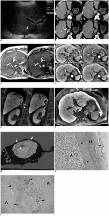
          We report a case of an adenoma arising from an ectopic adrenal gland mimicking a hepatocellular carcinoma in a heavy alcohol abuser. A MDCT showed a 2.7 low-attenuating nodule in segment VII of the liver through all CT phases. Compared to a precontrast image, however, a subtle enhancement was noted on the arterial phase CT image. On T1 weighted in- and opposed-phase MR images, an abundant fat component within the lesion was seen. Dynamic contrast-enhanced MR images after administration of gadolinium ethoxybenzyl diethylenetriaminepentaacetic acid (Gd-EOB-DTPA) more clearly depicted hypervascularity and wash-out of the lesion on arterial and portal phases, respectively. On delayed hepatobiliary phase MR images, obtained 20 minutes after Gd-EOB-DTPA administration, subtle uptake or retention of the contrast agent by the lesion was suspected. A tumorectomy was performed and adrenal adenoma from an ectopic adrenal gland within the liver was confirmed.

          Related collections

          Most cited references16

          • Record: found
          • Abstract: not found
          • Article: not found

          Management of hepatocellular carcinoma.

            Bookmark
            • Record: found
            • Abstract: found
            • Article: not found

            Adrenal masses: characterization with combined unenhanced and delayed enhanced CT.

            To assess the accuracy of a dedicated adrenal computed tomographic (CT) protocol. One hundred sixty-six adrenal masses were evaluated with a protocol consisting of unenhanced CT, and, for those with attenuation values greater than 10 HU, contrast material-enhanced and delayed enhanced CT. Attenuation values and enhancement washout calculations were obtained. An adenoma was diagnosed if a mass had an attenuation value of 10 HU or less at unenhanced CT or a percentage enhancement washout value of 60% or higher. The final diagnosis was adenoma in 127 masses and non-adenoma in 39. Masses measuring more than 10 HU on unenhanced CT scans were confirmed at biopsy (n = 28) or were examined for stability or change in size at follow-up CT performed at a minimum interval of 6 months (n = 33). Thirty-six (92%) of 39 non-adenomas and 124 (98%) of 127 adenomas were correctly characterized. The sensitivity and specificity of this protocol were 98% and 92%, respectively. This protocol correctly characterized 160 (96%) of 166 masses. With a combination of unenhanced and delayed enhanced CT, nearly all adrenal masses can be correctly categorized as adenomas or non-adenomas.
              Bookmark
              • Record: found
              • Abstract: found
              • Article: not found

              Adreno-hepatic fusion. An autopsy study.

              K-i Honma (1990)
              Adreno-hepatic fusion (AHF) is defined as adhesion of the liver and right adrenal cortex with close intermingling of the respective parenchymal cells. Contrary to the general opinion, AHF is a rather common event of unknown biological significance: AHF was exhibited by 63 of 636 unselected autopsy cases (9.9%). Its incidence is much higher in older age groups, suggesting that AHF may be an ageing phenomenon. No causative relationship was found to exist between occurrence of AHF and pathological conditions of both organs.
                Bookmark

                Author and article information

                Journal
                Korean J Radiol
                KJR
                Korean Journal of Radiology
                The Korean Society of Radiology
                1229-6929
                2005-8330
                Jan-Feb 2010
                28 December 2009
                : 11
                : 1
                : 126-130
                Affiliations
                [1 ]Department of Radiology, Seoul National University Hospital, Seoul 110-744, Korea.
                [2 ]Institute of Radiation Medicine, Seoul National University Hospital, Seoul 110-744, Korea.
                [3 ]Department of Pathology, Seoul National University Hospital, Seoul 110-744, Korea.
                Author notes
                Address reprint requests to: Se Hyung Kim, MD, Department of Radiology, Seoul National University Hospital, 101 Daehang-ro, Jongno-gu, Seoul 110-744, Korea. Tel. (822) 2072-2057, Fax. (822) 743-6385, shkim@ 123456radcom.snu.ac.kr
                Article
                10.3348/kjr.2010.11.1.126
                2799643
                20046505
                2d2e39c3-eeaf-40bf-a13a-2abb23cde798
                Copyright © 2010 The Korean Society of Radiology

                This is an Open Access article distributed under the terms of the Creative Commons Attribution Non-Commercial License ( http://creativecommons.org/licenses/by-nc/3.0) which permits unrestricted non-commercial use, distribution, and reproduction in any medium, provided the original work is properly cited.

                History
                : 03 May 2009
                : 26 August 2009
                Categories
                Case Report

                Radiology & Imaging
                ectopic adrenal gland,magnetic resonance (mr),adrenal adenoma,multi-detector computed tomography (mdct),liver

                Comments

                Comment on this article