3
views
0
recommends
+1 Recommend
0 collections
    0
    shares
      • Record: found
      • Abstract: found
      • Article: found
      Is Open Access

      Dysregulation of iron homeostasis and ferroptosis in sevoflurane and isoflurane associated perioperative neurocognitive disorders

      review-article

      Read this article at

      Bookmark
          There is no author summary for this article yet. Authors can add summaries to their articles on ScienceOpen to make them more accessible to a non-specialist audience.

          Abstract

          In recent years, sevoflurane and isoflurane are the most popular anesthetics in general anesthesia for their safe, rapid onset, and well tolerant. Nevertheless, many studies reported their neurotoxicity among pediatric and aged populations. This effect is usually manifested as cognitive impairment such as perioperative neurocognitive disorders. The wide application of sevoflurane and isoflurane during general anesthesia makes their safety a major health concern. Evidence indicates that iron dyshomeostasis and ferroptosis may establish a role in neurotoxicity of sevoflurane and isoflurane. However, the mechanisms of sevoflurane‐ and isoflurane‐induced neuronal injury were not fully understood, which poses a barrier to the treatment of its neurotoxicity. We, therefore, reviewed the current knowledge on mechanisms of iron dyshomeostasis and ferroptosis and aimed to promote a better understanding of their roles in sevoflurane‐ and isoflurane‐induced neurotoxicity.

          Abstract

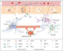
          Current knowledge on sevoflurane and isoflurane's effects on iron metabolism and ferroptosis in vulnerable brain. Sevoflurane and isoflurane anesthesia interrupt iron homeostasis and result in ferroptosis in brain. The expression of iron transporters including TFR1, FPN1, and DMT1 is impacted by sevoflurane anesthesia. Sevoflurane anesthesia causes iron overload via NMDAR‐RASD1‐DMT1 pathway‐mediated iron uptake. Iron overload induced by sevoflurane treatment can lead to Aβ accumulation, mitochondrial dysfunction, lipid ROS accumulation, and cognitive impairment in developmental or aging rodents. DFP and Fer‐1 can reverse these negative changes. SLC7A11‐GSH‐GPX4 axis is implicated into sevoflurane and isoflurane induced ferroptosis. Factors regulating this crucial axis are p‐Becline, MIB2, and ATF4. Echinatin inhibits sevoflurane‐induced ferroptosis by upregulatintg NRF2 pathway. In addition, mitochondria protective agents elamipretide (SS‐31), DMF and Mito‐Tempo treatment improved sevoflurane‐ and isoflurane‐induced cognitive deficits.

          Related collections

          Most cited references112

          • Record: found
          • Abstract: found
          • Article: not found

          Ferroptosis: an iron-dependent form of nonapoptotic cell death.

          Nonapoptotic forms of cell death may facilitate the selective elimination of some tumor cells or be activated in specific pathological states. The oncogenic RAS-selective lethal small molecule erastin triggers a unique iron-dependent form of nonapoptotic cell death that we term ferroptosis. Ferroptosis is dependent upon intracellular iron, but not other metals, and is morphologically, biochemically, and genetically distinct from apoptosis, necrosis, and autophagy. We identify the small molecule ferrostatin-1 as a potent inhibitor of ferroptosis in cancer cells and glutamate-induced cell death in organotypic rat brain slices, suggesting similarities between these two processes. Indeed, erastin, like glutamate, inhibits cystine uptake by the cystine/glutamate antiporter (system x(c)(-)), creating a void in the antioxidant defenses of the cell and ultimately leading to iron-dependent, oxidative death. Thus, activation of ferroptosis results in the nonapoptotic destruction of certain cancer cells, whereas inhibition of this process may protect organisms from neurodegeneration. Copyright © 2012 Elsevier Inc. All rights reserved.
            Bookmark
            • Record: found
            • Abstract: found
            • Article: not found

            Ferroptosis: A Regulated Cell Death Nexus Linking Metabolism, Redox Biology, and Disease

            Ferroptosis is a form of regulated cell death characterized by the iron-dependent accumulation of lipid hydroperoxides to lethal levels. Emerging evidence suggests that ferroptosis represents an ancient vulnerability caused by the incorporation of polyunsaturated fatty acids into cellular membranes, and cells have developed complex systems that exploit and defend against this vulnerability in different contexts. The sensitivity to ferroptosis is tightly linked to numerous biological processes, including amino acid, iron, and polyunsaturated fatty acid metabolism, and the biosynthesis of glutathione, phospholipids, NADPH, and coenzyme Q10. Ferroptosis has been implicated in the pathological cell death associated with degenerative diseases (i.e., Alzheimer's, Huntington's, and Parkinson's diseases), carcinogenesis, stroke, intracerebral hemorrhage, traumatic brain injury, ischemia-reperfusion injury, and kidney degeneration in mammals and is also implicated in heat stress in plants. Ferroptosis may also have a tumor-suppressor function that could be harnessed for cancer therapy. This Primer reviews the mechanisms underlying ferroptosis, highlights connections to other areas of biology and medicine, and recommends tools and guidelines for studying this emerging form of regulated cell death.
              Bookmark
              • Record: found
              • Abstract: found
              • Article: found
              Is Open Access

              Ferroptosis: past, present and future

              Ferroptosis is a new type of cell death that was discovered in recent years and is usually accompanied by a large amount of iron accumulation and lipid peroxidation during the cell death process; the occurrence of ferroptosis is iron-dependent. Ferroptosis-inducing factors can directly or indirectly affect glutathione peroxidase through different pathways, resulting in a decrease in antioxidant capacity and accumulation of lipid reactive oxygen species (ROS) in cells, ultimately leading to oxidative cell death. Recent studies have shown that ferroptosis is closely related to the pathophysiological processes of many diseases, such as tumors, nervous system diseases, ischemia-reperfusion injury, kidney injury, and blood diseases. How to intervene in the occurrence and development of related diseases by regulating cell ferroptosis has become a hotspot and focus of etiological research and treatment, but the functional changes and specific molecular mechanisms of ferroptosis still need to be further explored. This paper systematically summarizes the latest progress in ferroptosis research, with a focus on providing references for further understanding of its pathogenesis and for proposing new targets for the treatment of related diseases.
                Bookmark

                Author and article information

                Contributors
                mingyangsun1986@163.com
                zhangjiq@zzu.edu.cn
                Journal
                CNS Neurosci Ther
                CNS Neurosci Ther
                10.1111/(ISSN)1755-5949
                CNS
                CNS Neuroscience & Therapeutics
                John Wiley and Sons Inc. (Hoboken )
                1755-5930
                1755-5949
                09 February 2024
                February 2024
                : 30
                : 2 ( doiID: 10.1002/cns.v30.2 )
                : e14553
                Affiliations
                [ 1 ] Department of Anesthesiology and Perioperative medicine People's Hospital of Zhengzhou University, Henan Provincial People's Hospital, People's Hospital of Henan University Zhengzhou Henan Province China
                Author notes
                [*] [* ] Correspondence

                Mingyang Sun and Jiaqiang Zhang, Department of Anesthesia and Perioperative Medicine, Henan University People's Hospital, Zhengzhou University People's Hospital, Henan Provincial People's Hospital, Zhengzhou 450003, Henan Province, China.

                Email: mingyangsun1986@ 123456163.com and zhangjiq@ 123456zzu.edu.cn

                Author information
                https://orcid.org/0000-0002-6956-8599
                Article
                CNS14553 CNSNT-2023-094.R3
                10.1111/cns.14553
                10853900
                38334231
                2ccb3d59-41b5-4a6f-aeeb-a57bc408481a
                © 2023 The Authors. CNS Neuroscience & Therapeutics published by John Wiley & Sons Ltd.

                This is an open access article under the terms of the http://creativecommons.org/licenses/by/4.0/ License, which permits use, distribution and reproduction in any medium, provided the original work is properly cited.

                History
                : 06 November 2023
                : 19 January 2023
                : 22 November 2023
                Page count
                Figures: 4, Tables: 0, Pages: 11, Words: 7775
                Funding
                Funded by: the Natural Science Foundation of China
                Award ID: 82301448
                Award ID: 82071217
                Award ID: 82001147
                Funded by: Natural Science Foundation of Henan Province , doi 10.13039/501100006407;
                Award ID: 202300410353
                Categories
                Review
                Reviews
                Custom metadata
                2.0
                February 2024
                Converter:WILEY_ML3GV2_TO_JATSPMC version:6.3.6 mode:remove_FC converted:09.02.2024

                Neurosciences
                ferroptosis,iron homeostasis,isoflurane,neurotoxicity,sevoflurane
                Neurosciences
                ferroptosis, iron homeostasis, isoflurane, neurotoxicity, sevoflurane

                Comments

                Comment on this article