60
views
0
recommends
+1 Recommend
0 collections
    0
    shares
      • Record: found
      • Abstract: found
      • Article: not found

      Effects of Stimulants on Brain Function in Attention-Deficit/Hyperactivity Disorder: A Systematic Review and Meta-Analysis

      research-article

      Read this article at

      ScienceOpenPublisherPMC
      Bookmark
          There is no author summary for this article yet. Authors can add summaries to their articles on ScienceOpen to make them more accessible to a non-specialist audience.

          Abstract

          Background

          Psychostimulant medication, most commonly the catecholamine agonist methylphenidate, is the most effective treatment for attention-deficit/hyperactivity disorder (ADHD). However, relatively little is known on the mechanisms of action. Acute effects on brain function can elucidate underlying neurocognitive effects. We tested methylphenidate effects relative to placebo in functional magnetic resonance imaging (fMRI) during three disorder-relevant tasks in medication-naïve ADHD adolescents. In addition, we conducted a systematic review and meta-analysis of the fMRI findings of acute stimulant effects on ADHD brain function.

          Methods

          The fMRI study compared 20 adolescents with ADHD under either placebo or methylphenidate in a randomized controlled trial while performing stop, working memory, and time discrimination tasks. The meta-analysis was conducted searching PubMed, ScienceDirect, Web of Knowledge, Google Scholar, and Scopus databases. Peak coordinates of clusters of significant effects of stimulant medication relative to placebo or off medication were extracted for each study.

          Results

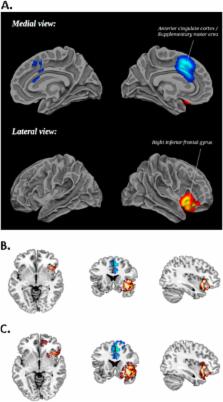
          The fMRI analysis showed that methylphenidate significantly enhanced activation in bilateral inferior frontal cortex (IFC)/insula during inhibition and time discrimination but had no effect on working memory networks. The meta-analysis, including 14 fMRI datasets and 212 children with ADHD, showed that stimulants most consistently enhanced right IFC/insula activation, which also remained for a subgroup analysis of methylphenidate effects alone. A more lenient threshold also revealed increased putamen activation.

          Conclusions

          Psychostimulants most consistently increase right IFC/insula activation, which are key areas of cognitive control and also the most replicated neurocognitive dysfunction in ADHD. These neurocognitive effects may underlie their positive clinical effects.

          Related collections

          Most cited references77

          • Record: found
          • Abstract: found
          • Article: not found

          Using Support Vector Machine to identify imaging biomarkers of neurological and psychiatric disease: a critical review.

          Standard univariate analysis of neuroimaging data has revealed a host of neuroanatomical and functional differences between healthy individuals and patients suffering a wide range of neurological and psychiatric disorders. Significant only at group level however these findings have had limited clinical translation, and recent attention has turned toward alternative forms of analysis, including Support-Vector-Machine (SVM). A type of machine learning, SVM allows categorisation of an individual's previously unseen data into a predefined group using a classification algorithm, developed on a training data set. In recent years, SVM has been successfully applied in the context of disease diagnosis, transition prediction and treatment prognosis, using both structural and functional neuroimaging data. Here we provide a brief overview of the method and review those studies that applied it to the investigation of Alzheimer's disease, schizophrenia, major depression, bipolar disorder, presymptomatic Huntington's disease, Parkinson's disease and autistic spectrum disorder. We conclude by discussing the main theoretical and practical challenges associated with the implementation of this method into the clinic and possible future directions. Copyright © 2012 Elsevier Ltd. All rights reserved.
            Bookmark
            • Record: found
            • Abstract: found
            • Article: not found

            Voxel-wise meta-analysis of grey matter changes in obsessive-compulsive disorder.

            Specific cortico-striato-thalamic circuits are hypothesised to mediate the symptoms of obsessive-compulsive disorder (OCD), but structural neuroimaging studies have been inconsistent. To conduct a meta-analysis of published and unpublished voxel-based morphometry studies in OCD. Twelve data-sets comprising 401 people with OCD and 376 healthy controls met inclusion criteria. A new improved voxel-based meta-analytic method, signed differential mapping (SDM), was developed to examine regions of increased and decreased grey matter volume in the OCD group v. control group. Results No between-group differences were found in global grey matter volumes. People with OCD had increased regional grey matter volumes in bilateral lenticular nuclei, extending to the caudate nuclei, as well as decreased volumes in bilateral dorsal medial frontal/anterior cingulate gyri. A descriptive analysis of quartiles, a sensitivity analysis as well as analyses of subgroups further confirmed these findings. Meta-regression analyses showed that studies that included individuals with more severe OCD were significantly more likely to report increased grey matter volumes in the basal ganglia. No effect of current antidepressant treatment was observed. Conclusions The results support a dorsal prefrontal-striatal model of the disorder and raise the question of whether functional alterations in other brain regions commonly associated with OCD, such as the orbitofrontal cortex, may reflect secondary compensatory strategies. Whether the reported differences between participants with OCD and controls precede the onset of the symptoms and whether they are specific to OCD remains to be established.
              Bookmark
              • Record: found
              • Abstract: not found
              • Article: not found

              Developmental trajectories of brain volume abnormalities in children and adolescents with attention-deficit/hyperactivity disorder.

              Various anatomic brain abnormalities have been reported for attention-deficit/hyperactivity disorder (ADHD), with varying methods, small samples, cross-sectional designs, and without accounting for stimulant drug exposure. To compare regional brain volumes at initial scan and their change over time in medicated and previously unmedicated male and female patients with ADHD and healthy controls. Case-control study conducted from 1991-2001 at the National Institute of Mental Health, Bethesda, Md, of 152 children and adolescents with ADHD (age range, 5-18 years) and 139 age- and sex-matched controls (age range, 4.5-19 years) recruited from the local community, who contributed 544 anatomic magnetic resonance images. Using completely automated methods, initial volumes and prospective age-related changes of total cerebrum, cerebellum, gray and white matter for the 4 major lobes, and caudate nucleus of the brain were compared in patients and controls. On initial scan, patients with ADHD had significantly smaller brain volumes in all regions, even after adjustment for significant covariates. This global difference was reflected in smaller total cerebral volumes (-3.2%, adjusted F(1,280) = 8.30, P =.004) and in significantly smaller cerebellar volumes (-3.5%, adjusted F(1,280) = 12.29, P =.001). Compared with controls, previously unmedicated children with ADHD demonstrated significantly smaller total cerebral volumes (overall F(2,288) = 6.65; all pairwise comparisons Bonferroni corrected, -5.8%; P =.002) and cerebellar volumes (-6.2%, F( 2,288) = 8.97, P<.001). Unmedicated children with ADHD also exhibited strikingly smaller total white matter volumes (F(2,288) = 11.65) compared with controls (-10.7%, P<.001) and with medicated children with ADHD (-8.9%, P<.001). Volumetric abnormalities persisted with age in total and regional cerebral measures (P =.002) and in the cerebellum (P =.003). Caudate nucleus volumes were initially abnormal for patients with ADHD (P =.05), but diagnostic differences disappeared as caudate volumes decreased for patients and controls during adolescence. Results were comparable for male and female patients on all measures. Frontal and temporal gray matter, caudate, and cerebellar volumes correlated significantly with parent- and clinician-rated severity measures within the ADHD sample (Pearson coefficients between -0.16 and -0.26; all P values were <.05). Developmental trajectories for all structures, except caudate, remain roughly parallel for patients and controls during childhood and adolescence, suggesting that genetic and/or early environmental influences on brain development in ADHD are fixed, nonprogressive, and unrelated to stimulant treatment.
                Bookmark

                Author and article information

                Contributors
                Journal
                Biol Psychiatry
                Biol. Psychiatry
                Biological Psychiatry
                Elsevier
                0006-3223
                1873-2402
                15 October 2014
                15 October 2014
                : 76
                : 8
                : 616-628
                Affiliations
                [a ]Departments of Child and Adolescent Psychiatry (KR, AAA, AIC, AS) and Neuroimaging (MJB), Institute of Psychiatry, King’s College London, United Kingdom
                [b ]Departments of Neuroimaging, Institute of Psychiatry, King’s College London, United Kingdom
                [c ]Fundació per a la Investigació i la Docència Maria Angustias Giménez Research Unit, Germanes Hospitalaries and Centro de Investigación Biomédica en Red de Salud Mental (JR), Barcelona, Spain
                Author notes
                [* ]Address correspondence to Katya Rubia, Ph.D., Institute of Psychiatry, Department of Child and Adolescent Psychiatry, King’s College London, SGDP, Po46, 16 De Crespigny Park, London SE5 8AF, United Kingdom katya.rubia@ 123456kcl.ac.uk
                Article
                S0006-3223(13)00952-9
                10.1016/j.biopsych.2013.10.016
                4183380
                24314347
                2bebe6b9-606e-4ffd-9c8b-28bcb5189b84
                © 2014 Society of Biological Psychiatry. All rights reserved.
                History
                : 19 June 2013
                : 17 October 2013
                : 18 October 2013
                Categories
                Archival Report

                Clinical Psychology & Psychiatry
                adhd,fmri,meta-analysis,methylphenidate,review,stimulants
                Clinical Psychology & Psychiatry
                adhd, fmri, meta-analysis, methylphenidate, review, stimulants

                Comments

                Comment on this article