36
views
0
recommends
+1 Recommend
0 collections
    0
    shares
      • Record: found
      • Abstract: found
      • Article: found
      Is Open Access

      Safety of Redo Hepatectomy for Colorectal Liver Metastases after Selective Interarterial Radiation Therapy: A Case Report

      case-report

      Read this article at

      Bookmark
          There is no author summary for this article yet. Authors can add summaries to their articles on ScienceOpen to make them more accessible to a non-specialist audience.

          Abstract

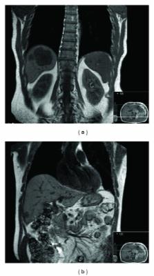
          Surgical resection is the only potentially curative strategy in the treatment of patients with colorectal liver metastases (CLM). Unfortunately, only about 10%–15% of patients are candidates for resection. Preoperative chemotherapy aims to increase the number of patients that may be eligible for liver resection by downsizing liver metastases. For patients with unresectable, chemotherapy refractory CLM the available treatment options are limited. Selective interarterial radiation therapy (SIRT) is one of the most promising treatment options for this group of patients. Although only a small number of these patients have been reported as becoming candidates for potentially curative hepatic resection following sufficient reduction in the volume of liver metastases, the question arises regarding the safety of liver resection in these patients. We report a case of a patient who presented unresectable liver relapse of CLM after previous right hepatectomy. He underwent SIRT which resulted in downsizing of the liver metastases making the patient candidate for left lateral sectionectomy. He underwent the redo hepatectomy without any complications. To the best of our knowledge, this is the first reported case of redo hepatectomy after SIRT for CLM.

          Related collections

          Most cited references41

          • Record: found
          • Abstract: found
          • Article: not found

          Trends in long-term survival following liver resection for hepatic colorectal metastases.

          To examine trends in outcomes of patients undergoing resection at a single tertiary care referral center over a 16-year period. Hepatic resection is considered the treatment of choice in selected patients with colorectal metastasis confined to the liver. Although a variety of retrospective studies have demonstrated improvements in short-term outcomes in recent years, changes in long-term survival over time are less well-established. Data from 226 consecutive patients undergoing potentially curative liver resection for colorectal metastases between 1984 and 1999 were analyzed. Actuarial survival rates related to prognostic determinants were analyzed using the log-rank test. The median survival for the entire cohort was 46 months, with 5- and 10-year survival rates of 40% and 26% respectively. Ninety-three patients operated on between 1984 and 1992 were found to have an overall survival of 31% at 5 years, compared to 58% for the 133 patients operated on during the more recent period (1993-1999). Both overall and disease-free survival were significantly better in the recent time period compared with the earlier period on both univariate and multivariate analyses. Other independent factors associated with improved survival included number of metastatic tumors < or = 3, negative resection margin, and CEA < 100. Comparisons were made between time periods for a variety of patient, tumor and treatment-related factors. Among all parameters studied, only resection type (anatomical versus nonanatomical), use of intraoperative ultrasonography, and perioperative chemotherapy administration differed between the early and recent time periods. Long-term survival following liver resection for colorectal metastases has improved significantly in recent years at our institution. Although the reasons for this survival trend are not clear, contributing factors may include the use of newer preoperative and intraoperative imaging, increased use of chemotherapy, and salvage surgical therapy.
            Bookmark
            • Record: found
            • Abstract: found
            • Article: not found

            Hepatic radiation toxicity: avoidance and amelioration.

            The refinement of radiation therapy and radioembolization techniques has led to a resurgent interest in radiation-induced liver disease (RILD). The awareness of technical and clinical parameters that influence the chance of RILD is important to guide patient selection and toxicity minimization strategies. "Classic" RILD is characterized by anicteric ascites and hepatomegaly and is unlikely to occur after a mean liver dose of approximately 30 Gy in conventional fractionation. By maintaining a low mean liver dose and sparing a "critical volume" of liver from radiation, stereotactic delivery techniques allow for the safe administration of higher tumor doses. Caution must be exercised for patients with hepatocellular carcinoma or pre-existing liver disease (eg, Child-Pugh score of B or C) because they are more susceptible to RILD that can manifest in a nonclassic pattern. Although no pharmacologic interventions have yet been proven to mitigate RILD, preclinical research shows the potential for therapies targeting transforming growth factor-β and for the transplantation of stem cells, hepatocytes, and liver progenitor cells as strategies that may restore liver function. Also, in the clinical setting of veno-occlusive liver disease after high-dose chemotherapy, agents with fibrinolytic and antithrombotic properties can reverse liver failure, suggesting a possible role in the setting of RILD.
              Bookmark
              • Record: found
              • Abstract: found
              • Article: not found

              Pathologic response and microdosimetry of (90)Y microspheres in man: review of four explanted whole livers.

              Radioactive microsphere (90)Y therapy is increasingly used for primary and metastatic solid tumors in the liver. We present an analysis of 4 explanted livers previously treated with (90)Y microsphere agents (glass or resin). One tumor nodule was analyzed with submillimeter three-dimensional microdosimetry. Four patients received hepatic artery delivery of (90)Y microspheres for unresectable hepatocellular and colon cancers. Whole livers were explanted as part of lifesaving cadaveric transplant in 2 patients with hepatoma. These patients had received glass microspheres as a procedural bridge to transplant. Autopsy was performed on 2 patients with colon cancer who died of progressive metastatic disease and who had been treated with resin microspheres. Complete pathologic review was performed on each whole liver, including estimation of the response of the tumor to therapy, distribution of microspheres in the tumor and normal liver tissues, and normal-tissue radiation response. A biopsy taken from the edge of a tumor nodule was sectioned serially for three-dimensional radiation dosimetry analyses. Three-dimensional microsphere coordinates within the biopsy specimen were used to calculate dosage using a three-dimensional dose kernel. Isodose coverage of tumor and normal liver areas and total dose delivered were determined. Preferential and heterogeneous deposition of microspheres was noted at the edge of tumor nodules compared with the center portion of the tumor or normal liver parenchyma. Both glass and resin microspheres delivered high cumulative doses to the tumor, which varied from 100 Gy to more than 3000 Gy. No veno-occlusive disease or widespread radiation hepatitis was seen. Microsphere ((90)Y) therapy delivers high numbers of spheres with resulting high total doses of radiation, preferentially in the periphery of tumors. Normal liver parenchyma showed little radiation effect away from the tumors. Heterogeneous high-dose regions in the tumor were produced by both glass and resin microspheres.
                Bookmark

                Author and article information

                Journal
                Case Rep Surg
                Case Rep Surg
                CRIS
                Case Reports in Surgery
                Hindawi Publishing Corporation
                2090-6900
                2090-6919
                2014
                4 March 2014
                : 2014
                : 712572
                Affiliations
                1Royal Marsden Hospital, Department of Academic Surgery, Upper GI/HPB Unit, Fulham Road, London SW3 6JJ, UK
                2Oncology, Imperial College Healthcare NHS Trust, Hammersmith Hospital, Du Cane Road, London W12 0HS, UK
                Author notes
                *Kyriakos Neofytou: kneophy2@ 123456gmail.com

                Academic Editors: N. A. Chowdri and H. Kawai

                Author information
                http://orcid.org/0000-0002-7937-4122
                Article
                10.1155/2014/712572
                3971541
                24716079
                2b9c87ea-b3ca-472e-a8a9-703efe4f9972
                Copyright © 2014 Kyriakos Neofytou et al.

                This is an open access article distributed under the Creative Commons Attribution License, which permits unrestricted use, distribution, and reproduction in any medium, provided the original work is properly cited.

                History
                : 28 November 2013
                : 19 January 2014
                Categories
                Case Report

                Surgery
                Surgery

                Comments

                Comment on this article