19
views
0
recommends
+1 Recommend
2 collections
    0
    shares
      • Record: found
      • Abstract: found
      • Article: found
      Is Open Access

      Natural killer cells associated with SARS-CoV-2 viral RNA shedding, antibody response and mortality in COVID-19 patients

      letter

      Read this article at

      Bookmark
          There is no author summary for this article yet. Authors can add summaries to their articles on ScienceOpen to make them more accessible to a non-specialist audience.

          Abstract

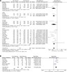
          Coronavirus disease 2019 (COVID-19) is a novel infectious viral disease caused by the severe acute respiratory syndrome coronavirus 2 (SARS-CoV-2). Two consecutively negative SARS-CoV-2 viral RNA test ( interval ≥ 24 hours), improved respiratory symptoms and obvious absorption of inflammation in pulmonary imaging are the discharge criteria for COVID-19 patients. The clearance profile of viral RNA in the upper respiratory tract specimens, including nasopharyngeal swab and/or oropharyngeal swabs, is related to innate immune cells such as Natural Killer cells. A total of 168 patients were included for the study. In this cohort, non-severe and severe groups showed significant differences in white blood cells, neutrophils, lymphocytes, basophils and platelets counts, as well as in infection related parameters such as CRP and serum cytokine IL-6. For lymphocyte subsets tests at admission, the severe group displayed significantly lower cell counts than the non-severe group. Higher counts of total T cells, CD4 + T cells, CD8 + T cells, and NK cells in peripheral blood showed a significant correlation with the shorter time taken to obtain the first negative viral RNA test and first positive IgM/ IgG antibody test. The number of B cells was only correlated with time to achieve the first positive IgM/IgG test. The count of NK cells was also correlated with a higher level of IgG antibody ( p = 0.025). The lymphocytopenia group had a significantly worse survival rate ( p = 0.022) and a longer duration ( p = 0.023) of viral shedding than the normal lymphocyte count group. A lower NK cell count correlates the most with the worse survival rate ( p<0.001) and a longer duration ( p<0.001) of viral shedding. This study suggests the potential value of allo-Natural Killer cell therapy as an universal COVID-19 treatment strategy.

          Related collections

          Most cited references13

          • Record: found
          • Abstract: found
          • Article: not found

          Clinical features of patients infected with 2019 novel coronavirus in Wuhan, China

          Summary Background A recent cluster of pneumonia cases in Wuhan, China, was caused by a novel betacoronavirus, the 2019 novel coronavirus (2019-nCoV). We report the epidemiological, clinical, laboratory, and radiological characteristics and treatment and clinical outcomes of these patients. Methods All patients with suspected 2019-nCoV were admitted to a designated hospital in Wuhan. We prospectively collected and analysed data on patients with laboratory-confirmed 2019-nCoV infection by real-time RT-PCR and next-generation sequencing. Data were obtained with standardised data collection forms shared by WHO and the International Severe Acute Respiratory and Emerging Infection Consortium from electronic medical records. Researchers also directly communicated with patients or their families to ascertain epidemiological and symptom data. Outcomes were also compared between patients who had been admitted to the intensive care unit (ICU) and those who had not. Findings By Jan 2, 2020, 41 admitted hospital patients had been identified as having laboratory-confirmed 2019-nCoV infection. Most of the infected patients were men (30 [73%] of 41); less than half had underlying diseases (13 [32%]), including diabetes (eight [20%]), hypertension (six [15%]), and cardiovascular disease (six [15%]). Median age was 49·0 years (IQR 41·0–58·0). 27 (66%) of 41 patients had been exposed to Huanan seafood market. One family cluster was found. Common symptoms at onset of illness were fever (40 [98%] of 41 patients), cough (31 [76%]), and myalgia or fatigue (18 [44%]); less common symptoms were sputum production (11 [28%] of 39), headache (three [8%] of 38), haemoptysis (two [5%] of 39), and diarrhoea (one [3%] of 38). Dyspnoea developed in 22 (55%) of 40 patients (median time from illness onset to dyspnoea 8·0 days [IQR 5·0–13·0]). 26 (63%) of 41 patients had lymphopenia. All 41 patients had pneumonia with abnormal findings on chest CT. Complications included acute respiratory distress syndrome (12 [29%]), RNAaemia (six [15%]), acute cardiac injury (five [12%]) and secondary infection (four [10%]). 13 (32%) patients were admitted to an ICU and six (15%) died. Compared with non-ICU patients, ICU patients had higher plasma levels of IL2, IL7, IL10, GSCF, IP10, MCP1, MIP1A, and TNFα. Interpretation The 2019-nCoV infection caused clusters of severe respiratory illness similar to severe acute respiratory syndrome coronavirus and was associated with ICU admission and high mortality. Major gaps in our knowledge of the origin, epidemiology, duration of human transmission, and clinical spectrum of disease need fulfilment by future studies. Funding Ministry of Science and Technology, Chinese Academy of Medical Sciences, National Natural Science Foundation of China, and Beijing Municipal Science and Technology Commission.
            Bookmark
            • Record: found
            • Abstract: found
            • Article: not found

            Virological assessment of hospitalized patients with COVID-2019

            Coronavirus disease 2019 (COVID-19) is an acute infection of the respiratory tract that emerged in late 20191,2. Initial outbreaks in China involved 13.8% of cases with severe courses, and 6.1% of cases with critical courses3. This severe presentation may result from the virus using a virus receptor that is expressed predominantly in the lung2,4; the same receptor tropism is thought to have determined the pathogenicity-but also aided in the control-of severe acute respiratory syndrome (SARS) in 20035. However, there are reports of cases of COVID-19 in which the patient shows mild upper respiratory tract symptoms, which suggests the potential for pre- or oligosymptomatic transmission6-8. There is an urgent need for information on virus replication, immunity and infectivity in specific sites of the body. Here we report a detailed virological analysis of nine cases of COVID-19 that provides proof of active virus replication in tissues of the upper respiratory tract. Pharyngeal virus shedding was very high during the first week of symptoms, with a peak at 7.11 × 108 RNA copies per throat swab on day 4. Infectious virus was readily isolated from samples derived from the throat or lung, but not from stool samples-in spite of high concentrations of virus RNA. Blood and urine samples never yielded virus. Active replication in the throat was confirmed by the presence of viral replicative RNA intermediates in the throat samples. We consistently detected sequence-distinct virus populations in throat and lung samples from one patient, proving independent replication. The shedding of viral RNA from sputum outlasted the end of symptoms. Seroconversion occurred after 7 days in 50% of patients (and by day 14 in all patients), but was not followed by a rapid decline in viral load. COVID-19 can present as a mild illness of the upper respiratory tract. The confirmation of active virus replication in the upper respiratory tract has implications for the containment of COVID-19.
              Bookmark
              • Record: found
              • Abstract: found
              • Article: found
              Is Open Access

              Lymphopenia in severe coronavirus disease-2019 (COVID-19): systematic review and meta-analysis

              Objective Clinical and laboratory biomarkers to predict the severity of coronavirus disease 2019 (COVID-19) are essential in this pandemic situation of which resource allocation must be urgently prepared especially in the context of respiratory support readiness. Lymphocyte count has been a marker of interest since the first COVID-19 publication. We conducted a systematic review and meta-analysis in order to investigate the association of lymphocyte count on admission and the severity of COVID-19. We would also like to analyze whether patient characteristics such as age and comorbidities affect the relationship between lymphocyte count and COVID-19. Methods Comprehensive and systematic literature search was performed from PubMed, SCOPUS, EuropePMC, ProQuest, Cochrane Central Databases, and Google Scholar. Research articles in adult patients diagnosed with COVID-19 with information on lymphocyte count and several outcomes of interest, including mortality, acute respiratory distress syndrome (ARDS), intensive care unit (ICU) care, and severe COVID-19, were included in the analysis. Inverse variance method was used to obtain mean differences and its standard deviations. Maentel-Haenszel formula was used to calculate dichotomous variables to obtain odds ratios (ORs) along with its 95% confidence intervals. Random-effect models were used for meta-analysis regardless of heterogeneity. Restricted-maximum likelihood random-effects meta-regression was performed for age, gender, cardiac comorbidity, hypertension, diabetes mellitus, COPD, and smoking. Results There were a total of 3099 patients from 24 studies. Meta-analysis showed that patients with poor outcome have a lower lymphocyte count (mean difference − 361.06 μL [− 439.18, − 282.95], p < 0.001; I 2 84%) compared to those with good outcome. Subgroup analysis showed lower lymphocyte count in patients who died (mean difference − 395.35 μL [− 165.64, − 625.07], p < 0.001; I 2 87%), experienced ARDS (mean difference − 377.56 μL [− 271.89, − 483.22], p < 0.001; I 2 0%), received ICU care (mean difference − 376.53 μL [− 682.84, − 70.22], p = 0.02; I 2 89%), and have severe COVID-19 (mean difference − 353.34 μL [− 250.94, − 455.73], p < 0.001; I 2 85%). Lymphopenia was associated with severe COVID-19 (OR 3.70 [2.44, 5.63], p < 0.001; I 2 40%). Meta-regression showed that the association between lymphocyte count and composite poor outcome was affected by age (p = 0.034). Conclusion This meta-analysis showed that lymphopenia on admission was associated with poor outcome in patients with COVID-19.
                Bookmark

                Author and article information

                Contributors
                ken.young@duke.edu
                qianwb@zju.edu.cn
                Journal
                Exp Hematol Oncol
                Exp Hematol Oncol
                Experimental Hematology & Oncology
                BioMed Central (London )
                2162-3619
                27 January 2021
                27 January 2021
                2021
                : 10
                : 5
                Affiliations
                [1 ]GRID grid.13402.34, ISNI 0000 0004 1759 700X, Department of Hematology, The Second Affiliated Hospital, , Zhejiang University School of Medicine, ; 310009 Hangzhou, China
                [2 ]GRID grid.73113.37, ISNI 0000 0004 0369 1660, The Third Affiliated Hospital of Naval Medical University, ; 200438 Shanghai, China
                [3 ]Wuhan Huoshenshan Hospital, 430100 Wuhan, China
                [4 ]GRID grid.13402.34, ISNI 0000 0004 1759 700X, Department of Intensive Care Unit, The Second Affiliated Hospital, , Zhejiang University School of Medicine, ; 310009 Hangzhou, China
                [5 ]GRID grid.13402.34, ISNI 0000 0004 1759 700X, Department of Dentist, The Second Affiliated Hospital, , Zhejiang University School of Medicine, ; 310009 Hangzhou, China
                [6 ]GRID grid.189509.c, ISNI 0000000100241216, Hematopathology Division, Department of Pathology, , Duke University Medical Center, Duke University Cancer Center, ; Durham, NC USA
                [7 ]GRID grid.429222.d, ISNI 0000 0004 1798 0228, National Clinical Research Center for Hematologic Diseases, , The First Affiliated Hospital of Soochow University, ; Jiangsu 215006 Suzhou, China
                Author information
                http://orcid.org/0000-0002-9817-6674
                Article
                199
                10.1186/s40164-021-00199-1
                7839286
                33504359
                2ab99102-db3a-45d8-bebb-8a47df430b0f
                © The Author(s) 2021

                Open AccessThis article is licensed under a Creative Commons Attribution 4.0 International License, which permits use, sharing, adaptation, distribution and reproduction in any medium or format, as long as you give appropriate credit to the original author(s) and the source, provide a link to the Creative Commons licence, and indicate if changes were made. The images or other third party material in this article are included in the article's Creative Commons licence, unless indicated otherwise in a credit line to the material. If material is not included in the article's Creative Commons licence and your intended use is not permitted by statutory regulation or exceeds the permitted use, you will need to obtain permission directly from the copyright holder. To view a copy of this licence, visit http://creativecommons.org/licenses/by/4.0/. The Creative Commons Public Domain Dedication waiver ( http://creativecommons.org/publicdomain/zero/1.0/) applies to the data made available in this article, unless otherwise stated in a credit line to the data.

                History
                : 17 November 2020
                : 4 January 2021
                Funding
                Funded by: The Translation Research Grant of NCRCH
                Award ID: 2020ZKZC01
                Categories
                Letter to the Editor
                Custom metadata
                © The Author(s) 2021

                Oncology & Radiotherapy
                covid-19,sars-cov-2,natural killer cell,rna viral shedding,antibody response

                Comments

                Comment on this article