116
views
0
recommends
+1 Recommend
0 collections
    0
    shares
      • Record: found
      • Abstract: found
      • Article: found
      Is Open Access

      α-Solanine induces ROS-mediated autophagy through activation of endoplasmic reticulum stress and inhibition of Akt/mTOR pathway

      research-article

      Read this article at

      Bookmark
          There is no author summary for this article yet. Authors can add summaries to their articles on ScienceOpen to make them more accessible to a non-specialist audience.

          Abstract

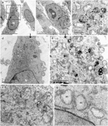
          α-Solanine is a glycoalkaloid found in species of the nightshade family including potato. It was primarily reported to have toxic effects in humans. However, there is a growing body of literature demonstrating in vitro and in vivo anticancer activity of α-solanine. Most of these studies have shown activation of apoptosis as the underlying mechanism in antitumor activity of α-solanine. In this study, we report α-solanine as a potential inducer of autophagy, which may act synergistically or in parallel with apoptosis to exert its cytotoxic effect. Induction of autophagy was demonstrated by several assays including electron microscopy, immunoblotting of autophagy markers and immunofluorescence for LC3 (microtubule-associated protein 1 (MAP1) light chain-3) puncta. α-Solanine-induced autophagic flux was demonstrated by additionally enhanced – turnover of LC3-II and – accumulation of LC3-specific puncta after co-incubation of cells with either of the autophagolysosome inhibitors – chloroquine and – bafilomycin A1. We also demonstrated α-solanine-induced oxidative damage in regulating autophagy where pre-incubation of cells with reactive oxygen species (ROS) scavenger resulted in suppression of CM-H 2DCFDA (5 (and 6)-chloromethyl-2′,7′-dichlorodihydrofluorescein diacetate acetyl ester) fluorescence as well as decrease in LC3-II turnover. α-Solanine treatment caused an increase in the expression of endoplasmic reticulum (ER) stress proteins (BiP, activating transcription factor 6 (ATF6), X-box-binding protein 1, PERK, inositol-requiring transmembrane kinase/endonuclease 1, ATF4 and CCAAT-enhancer-binding protein (C/EBP)-homologous protein) suggesting activation of unfolded protein response pathway. Moreover, we found downregulation of phosphorylated Akt (Thr 308 and Ser 473), mammalian target of rapamycin (mTOR; Ser 2448 and Ser 2481) and 4E-BP1 (Thr 37/46) by α-solanine implying suppression of the Akt/mTOR pathway. Collectively, our results signify that α-solanine induces autophagy to exert anti-proliferative activity by triggering ER stress and inhibiting Akt/mTOR signaling pathway.

          Related collections

          Most cited references36

          • Record: found
          • Abstract: found
          • Article: not found

          Endoplasmic reticulum stress: cell life and death decisions.

          C. Xu (2005)
          Disturbances in the normal functions of the ER lead to an evolutionarily conserved cell stress response, the unfolded protein response, which is aimed initially at compensating for damage but can eventually trigger cell death if ER dysfunction is severe or prolonged. The mechanisms by which ER stress leads to cell death remain enigmatic, with multiple potential participants described but little clarity about which specific death effectors dominate in particular cellular contexts. Important roles for ER-initiated cell death pathways have been recognized for several diseases, including hypoxia, ischemia/reperfusion injury, neurodegeneration, heart disease, and diabetes.
            Bookmark
            • Record: found
            • Abstract: found
            • Article: not found

            Autophagy is activated for cell survival after endoplasmic reticulum stress.

            Eukaryotic cells deal with accumulation of unfolded proteins in the endoplasmic reticulum (ER) by the unfolded protein response, involving the induction of molecular chaperones, translational attenuation, and ER-associated degradation, to prevent cell death. Here, we found that the autophagy system is activated as a novel signaling pathway in response to ER stress. Treatment of SK-N-SH neuroblastoma cells with ER stressors markedly induced the formation of autophagosomes, which were recognized at the ultrastructural level. The formation of green fluorescent protein (GFP)-LC3-labeled structures (GFP-LC3 "dots"), representing autophagosomes, was extensively induced in cells exposed to ER stress with conversion from LC3-I to LC3-II. In IRE1-deficient cells or cells treated with c-Jun N-terminal kinase (JNK) inhibitor, the autophagy induced by ER stress was inhibited, indicating that the IRE1-JNK pathway is required for autophagy activation after ER stress. In contrast, PERK-deficient cells and ATF6 knockdown cells showed that autophagy was induced after ER stress in a manner similar to the wild-type cells. Disturbance of autophagy rendered cells vulnerable to ER stress, suggesting that autophagy plays important roles in cell survival after ER stress.
              Bookmark
              • Record: found
              • Abstract: found
              • Article: not found

              Lysosomes and autophagy in cell death control.

              Lysosomal hydrolases participate in the digestion of endocytosed and autophagocytosed material inside the lysosomal/autolysosomal compartment in acute cell death when released into the cytosol and in cancer progression following their release into the extracellular space. Lysosomal alterations are common in cancer cells. The increased expression and altered trafficking of lysosomal enzymes participates in tissue invasion, angiogenesis and sensitization to the lysosomal death pathway. But lysosomal heat-shock protein 70 locally prevents lysosomal-membrane permeabilization. Similarly, alterations in the autophagic compartment are linked to carcinogenesis and resistance to chemotherapy. Targeting these pathways might constitute a novel approach to cancer therapy.
                Bookmark

                Author and article information

                Journal
                Cell Death Dis
                Cell Death Dis
                Cell Death & Disease
                Nature Publishing Group
                2041-4889
                August 2015
                27 August 2015
                1 August 2015
                : 6
                : 8
                : e1860
                Affiliations
                [1 ]Biochemistry Division, CSIR-Central Drug Research Institute , Lucknow, India
                [2 ]Electron Microscopy Unit, CSIR-Central Drug Research Institute , Lucknow, India
                [3 ]Sophisticated Analytical Instruments Facilities, CSIR-Central Drug Research Institute , Lucknow, India
                [4 ]Academy of Scientific and Innovative Research , Chennai, India
                Author notes
                [* ]Electron Microscopy Unit, CSIR-Central Drug Research Institute, Sector-10, Jankipuram Extension , Lucknow 226 031, India. Tel: +91 522 2772450; Fax: +91 522 2771941; E-mail: k_mitra@ 123456cdri.res.in
                [* ]Biochemistry Division, CSIR-Central Drug Research Institute, Sector-10, Jankipuram Extension , Lucknow 226 031, India. Tel: +91 522 2772450; Fax: +91 522 2771941; E-mail: j_sarkar@ 123456cdri.res.in or jayantavirol@ 123456gmail.com
                [5]

                These authors contributed equally to this work.

                Article
                cddis2015219
                10.1038/cddis.2015.219
                4558510
                26313911
                29eabd43-a56c-4ca4-b870-83053c706a80
                Copyright © 2015 Macmillan Publishers Limited

                Cell Death and Disease is an open-access journal published by Nature Publishing Group. This work is licensed under a Creative Commons Attribution 4.0 International License. The images or other third party material in this article are included in the article's Creative Commons license, unless indicated otherwise in the credit line; if the material is not included under the Creative Commons license, users will need to obtain permission from the license holder to reproduce the material. To view a copy of this license, visit http://creativecommons.org/licenses/by/4.0/

                History
                : 10 February 2015
                : 09 June 2015
                : 19 June 2015
                Categories
                Original Article

                Cell biology
                Cell biology

                Comments

                Comment on this article