15
views
0
recommends
+1 Recommend
0 collections
    0
    shares
      • Record: found
      • Abstract: found
      • Article: found
      Is Open Access

      Parkinsonism: a rare manifestation of craniopharyngioma

      case-report

      Read this article at

      Bookmark
          There is no author summary for this article yet. Authors can add summaries to their articles on ScienceOpen to make them more accessible to a non-specialist audience.

          Abstract

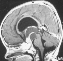
          Craniopharyngioma is a non-glial, non-malignant intracranial tumor of ectodermal origin, which arises from a remnant of Rathke’s pouch. This tumor accounts for 5.6 to 13% of intracranial tumors in children. This paper discusses a case of craniopharyngioma in a five-year-old boy. An MRI scan of his brain showed a huge sella and supra sella cystic-solid lesion that had invaded the prepontine and interpeduncular cisterns, filling of 3rd ventricle and hydrocephalus. The patient operated via interhemispheric subfrontal through lamina terminalis and the tumor dissected from all part of brain stem and total resection achieved. After surgery Parkinsonism was worse for 3 days and levodopa started for 3 days. Parkinsonism was gone and after one week levodopa discontinued. This case practically implied that decompression of mass effect of tumor on brain stem and short-term management with levodopa can improve Parkinsonism due to midline compressive brain tumors without basal ganglia involvement.

          Related collections

          Most cited references15

          • Record: found
          • Abstract: found
          • Article: found
          Is Open Access

          Long Term Sequelae of Pediatric Craniopharyngioma – Literature Review and 20 Years of Experience

          Craniopharyngioma are rare histologically benign brain tumors that develop in the pituitary–hypothalamic area. They may invade nearby anatomical structures causing significant rates of neurological, neurocognitive, and endocrinological complications including remarkable hypothalamic damage. Information regarding long term implications of the tumors and treatment in the pediatric population is accumulating, and treatment goals appear to be changing accordingly. In this review we aim to present data regarding long term complications of craniopharyngioma in children and adolescents and our experience from a large tertiary center. Hypothalamic dysfunction was noted to be the most significant complication, adversely affecting quality of life in survivors. Obesity, fatigue, and sleep disorders are the most notable manifestations of this dysfunction, and treatment is extremely difficult. Changes in management in recent years show a potential for improved long term outcomes; we found a trend toward less aggressive surgical management and increasing use of adjuvant treatment, accompanied by a decrease in complication rates.
            Bookmark
            • Record: found
            • Abstract: found
            • Article: found
            Is Open Access

            Diagnostics, Treatment, and Follow-Up in Craniopharyngioma

            Craniopharyngiomas are partly cystic embryogenic malformations of the sellar and parasellar region, with up to half the 0.5–2.0 new cases per million population per year occur in children and adolescents. Diagnosis profile for pediatric and adult craniopharyngioma is characterized by a combination of headache, visual impairment, and polyuria/polydipsia, which can also include significant weight gain. In children, growth retardation, and/or premature puberty often occur later or postoperatively. Recommended therapy with favorable tumor localization is complete resection; with unfavorable tumor localization (optic nerve and/or hypothalamic involvement), consensus is still pending whether a limited resection followed by local irradiation is more prudent. Even though overall survival rates are high (92%), recurrences after complete resection and progressions after incomplete resection can be expected. Accordingly, a randomized multinational trial (KRANIOPHARYNGEOM 2007) has been established to identify optimal diagnosis, treatment (particularly the ideal time point of irradiation after incomplete resection), and quality of life strategies of this chronic disease – most notably the morbid hypothalamic obesity in ∼50% of long-term survivors. We report on craniopharyngioma origins, its pathological manifestations, and specific challenges these sequelae pose regarding diagnosis, treatment, and life-long multi-discipline quality of life management for both adult and childhood craniopharyngioma patients.
              Bookmark
              • Record: found
              • Abstract: found
              • Article: not found

              Movement disorders caused by brain tumours.

              Movement disorders are uncommon presenting features of brain tumours. Early recognition of such lesions is important to arrest further deficit. We treated seven patients with movement disorders secondary to brain tumours over a period of seven years. Only two of these were intrinsic thalamic tumours (astrocytomas) while the rest were extrinsic tumours. The intrinsic tumours were accompanied by hemichorea. Among the extrinsic tumours, there was one pituitary macroadenoma with hemiballismus and four meningiomas with parkinsonism. Symptoms were unilateral in all patients except one with anterior third falcine meningioma who had bilateral rest tremors. There was relief in movement disorders observed after surgery. Imaging by computed tomography or magnetic resonance imaging is mandatory in the evaluation of movement disorders, especially if the presentation is atypical, unilateral and/or accompanied by long tract signs.
                Bookmark

                Author and article information

                Journal
                Electron Physician
                Electron Physician
                Electronic physician
                Electronic Physician
                Electronic physician
                2008-5842
                Apr-Jun 2015
                05 June 2015
                : 7
                : 2
                : 1027-1031
                Affiliations
                [1 ]Assistant Professor, Neurosurgery ward of Rasool-e-Akram Hospital Complex, Department of Neurosurgery, Iran University of Medical Science, Tehran, Iran
                [2 ]Associate Professor, Neurosurgery ward of Rasool-e-Akram Hospital Complex, Department of Neurosurgery, Iran University of Medical Science, Tehran, Iran
                [3 ]Medical Doctor, Resident of Neurosurgery, Neurosurgery ward of Rasool-e-Akram Hospital Complex, Department of Neurosurgery, Iran University of Medical Science, Tehran, Iran
                Author notes
                Corresponding author: Dr. Hossein Ghalaenovi, Neurosurgery ward of Rasool-e-Akram Hospital Complex, Department of Neurosurgery, Iran University of Medical Sciences, Tehran, Iran. Tel: +98.9124623806, Fax: +98.64352228. Email: hghalaenovi@ 123456yahoo.com
                Article
                epj-07-1027
                10.14661/2015.1027-1031
                4477761
                26120410
                299cad72-89d0-4737-abf2-415cae8b2553
                © 2015 The Authors

                This is an open access article under the terms of the Creative Commons Attribution-NonCommercial-NoDerivs License, which permits use and distribution in any medium, provided the original work is properly cited, the use is non-commercial and no modifications or adaptations are made.

                History
                : 01 February 2015
                : 26 April 2015
                Categories
                Case Report

                craniopharyngioma,parkinsonism,brain stem
                craniopharyngioma, parkinsonism, brain stem

                Comments

                Comment on this article