10
views
0
recommends
+1 Recommend
0 collections
    0
    shares
      • Record: found
      • Abstract: found
      • Article: found
      Is Open Access

      Root and Canal Morphology of Mandibular Premolars in a Saudi Subpopulation: A Cone-Beam Computed Tomography Study

      research-article

      Read this article at

      Bookmark
          There is no author summary for this article yet. Authors can add summaries to their articles on ScienceOpen to make them more accessible to a non-specialist audience.

          Abstract

          Objectives

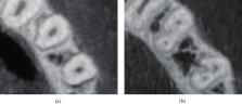
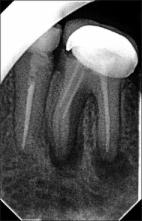
          The efficacy of root canal therapy is dependent on a thorough understanding of both normal and aberrant root canal morphology. As a result, the purpose of this study was to use CBCT to characterize the exact root and canal morphology of mandibular premolars in a Saudi subpopulation.

          Methods

          The current study included 1000 mandibular premolars (507 first premolars and 493 second premolars) with completely developed roots. CBCT was performed to assess the shape of the roots and to classify the canal anatomy according to Vertucci's classification. The incidence and similarity of the left and right sides, as well as men and women, were investigated. The data were examined using the chi-square test.

          Results

          Of the 507 mandibular first premolars analyzed, 484 (95.5%) had one root, whereas 23 (4.5%) had two roots. Of the 493 mandibular second premolars analyzed, 489 (99.2%) had one root, whereas four teeth had two roots (0.8%). There were no statistically significant variations in the number of roots identified across groups ( p > 0.05). The most prevalent in mandibular first premolars was type I, accounting for 70.0% ( n = 355) of the studied sample, followed by type II (14.2%, n = 72) and type IV (10.1%, n = 51). For mandibular second premolar, type I had the highest incidence (449 (91.1%)), followed by type II (5.7%, n = 28).

          Conclusion

          In a Saudi subpopulation, the majority of mandibular first and second premolar teeth had a single root with a type I canal system. On the other hand, numerous roots with various canal classifications were found.

          Related collections

          Most cited references51

          • Record: found
          • Abstract: found
          • Article: not found

          Root canal anatomy of the human permanent teeth.

          Two thousand four hundred human permanent teeth were decalcified, injected with dye, and cleared in order to determine the number of root canals and their different types, the ramifications of the main root canals, the location of apical foramina and transverse anastomoses, and the frequency of apical deltas.
            Bookmark
            • Record: found
            • Abstract: not found
            • Article: not found

            Microbiologic analysis of teeth with failed endodontic treatment and the outcome of conservative re-treatment

              Bookmark
              • Record: found
              • Abstract: found
              • Article: found
              Is Open Access

              Failure of endodontic treatment: The usual suspects

              Inappropriate mechanical debridement, persistence of bacteria in the canals and apex, poor obturation quality, over and under extension of the root canal filling, and coronal leakage are some of the commonly attributable causes of failure. Despite the high success rate of endodontic treatment, failures do occur in a large number of cases and most of the times can be attributed to the already stated causes. With an ever increasing number of endodontic treatments being done each day, it has become imperative to avoid or minimize the most fundamental of reasons leading to endodontic failure. This paper reviews the most common causes of endodontic failure along with radiographic examples.
                Bookmark

                Author and article information

                Contributors
                Journal
                Int J Dent
                Int J Dent
                ijd
                International Journal of Dentistry
                Hindawi
                1687-8728
                1687-8736
                2022
                8 March 2022
                : 2022
                : 4038909
                Affiliations
                1Department of Restorative Dental Science, University of Ha'il, Ha'il, Saudi Arabia
                2Ministry of Health, Ha'il, Saudi Arabia
                Author notes

                Academic Editor: Luca Testarelli

                Author information
                https://orcid.org/0000-0002-8865-6826
                https://orcid.org/0000-0003-0918-112X
                https://orcid.org/0000-0002-6647-7368
                https://orcid.org/0000-0001-6980-1656
                https://orcid.org/0000-0002-9495-9444
                https://orcid.org/0000-0001-9507-5416
                https://orcid.org/0000-0001-6124-0129
                Article
                10.1155/2022/4038909
                8924601
                35310460
                293a2d73-098b-48c1-ae8f-41f53adbefa0
                Copyright © 2022 Saad M. Al-Zubaidi et al.

                This is an open access article distributed under the Creative Commons Attribution License, which permits unrestricted use, distribution, and reproduction in any medium, provided the original work is properly cited.

                History
                : 6 November 2021
                : 3 January 2022
                : 11 February 2022
                Categories
                Research Article

                Dentistry
                Dentistry

                Comments

                Comment on this article