3
views
0
recommends
+1 Recommend
0 collections
    0
    shares
      • Record: found
      • Abstract: found
      • Article: found
      Is Open Access

      The Space between the Pial Sheath and the Cortical Venous Wall May Connect to the Meningeal Lymphatics

      brief-report

      Read this article at

      Bookmark
          There is no author summary for this article yet. Authors can add summaries to their articles on ScienceOpen to make them more accessible to a non-specialist audience.

          Abstract

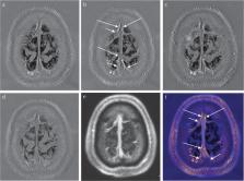
          We currently obtain pre- and post-contrast enhanced whole brain 3D-real inversion recovery images for the evaluation of endolymphatic hydrops. We noticed that the space between the pial sheath surrounding the cortical veins and the cortical venous wall is enhanced and this enhancement seems to connect to the meningeal lymphatics along superior sagittal sinus. This new anatomical concept regarding the outflow from the glymphatic system might be important for the future research in neuroscience.

          Related collections

          Most cited references7

          • Record: found
          • Abstract: found
          • Article: found
          Is Open Access

          Human and nonhuman primate meninges harbor lymphatic vessels that can be visualized noninvasively by MRI

          Here, we report the existence of meningeal lymphatic vessels in human and nonhuman primates (common marmoset monkeys) and the feasibility of noninvasively imaging and mapping them in vivo with high-resolution, clinical MRI. On T2-FLAIR and T1-weighted black-blood imaging, lymphatic vessels enhance with gadobutrol, a gadolinium-based contrast agent with high propensity to extravasate across a permeable capillary endothelial barrier, but not with gadofosveset, a blood-pool contrast agent. The topography of these vessels, running alongside dural venous sinuses, recapitulates the meningeal lymphatic system of rodents. In primates, meningeal lymphatics display a typical panel of lymphatic endothelial markers by immunohistochemistry. This discovery holds promise for better understanding the normal physiology of lymphatic drainage from the central nervous system and potential aberrations in neurological diseases.
            Bookmark
            • Record: found
            • Abstract: found
            • Article: not found

            Interrelationships of the pia mater and the perivascular (Virchow-Robin) spaces in the human cerebrum.

            Biopsies of histologically normal adult human cerebral cortex, underlying white matter and overlying leptomeninges were taken from frontal and temporal lobectomy specimens excised during the removal of cerebral tumours. Multiple blocks from 6 patients (aged 18-53 years) were examined by light and transmission electron microscopy. A thin sheath of pia mater cells was found to surround completely arterioles and arteries in the brain, in the subpial space and in the subarachnoid space. Pia mater cells, forming the perivascular sheath, were identified by the presence of desmosomes or small nexus junctions and by continuity with the pia mater itself. The presence of the pial sheath suggests that the perivascular spaces around intracerebral arteries are in direct continuity with the perivascular spaces around subarachnoid arteries. No similar pial sheath was observed around intracerebral or subpial venules. The role of the periarterial spaces, enclosed by the pial sheath, is discussed in relation to the results of physiological experiments suggesting drainage of interstitial fluid from brain tissue into the perivascular pathways along major cerebral arteries in the subarachnoid space. As arterioles in the brain become smaller and lose their smooth muscle coats, the pial sheath becomes incomplete. The anatomical relationships between the pia mater and blood vessels in the human cerebrum is summarised diagrammatically, and a possible role for pial cells as an enzymic barrier protecting the brain from exogenous catecholamines is discussed.
              Bookmark
              • Record: found
              • Abstract: found
              • Article: not found

              Where are we? The anatomy of the murine cortical meninges revisited for intravital imaging, immunology, and clearance of waste from the brain.

              Rapid progress is being made in understanding the roles of the cerebral meninges in the maintenance of normal brain function, in immune surveillance, and as a site of disease. Most basic research on the meninges and the neural brain is now done on mice, major attractions being the availability of reporter mice with fluorescent cells, and of a huge range of antibodies useful for immunocytochemistry and the characterization of isolated cells. In addition, two-photon microscopy through the unperforated calvaria allows intravital imaging of the undisturbed meninges with sub-micron resolution. The anatomy of the dorsal meninges of the mouse (and, indeed, of all mammals) differs considerably from that shown in many published diagrams: over cortical convexities, the outer layer, the dura, is usually thicker than the inner layer, the leptomeninx, and both layers are richly vascularized and innervated, and communicate with the lymphatic system. A membrane barrier separates them and, in disease, inflammation can be localized to one layer or the other, so experimentalists must be able to identify the compartment they are studying. Here, we present current knowledge of the functional anatomy of the meninges, particularly as it appears in intravital imaging, and review their role as a gateway between the brain, blood, and lymphatics, drawing on information that is scattered among works on different pathologies.
                Bookmark

                Author and article information

                Journal
                Magn Reson Med Sci
                Magn Reson Med Sci
                mrms
                Magnetic Resonance in Medical Sciences
                Japanese Society for Magnetic Resonance in Medicine
                1347-3182
                1880-2206
                2020
                15 October 2019
                : 19
                : 1
                : 1-4
                Affiliations
                [1 ]Department of Radiology, Nagoya University Graduate School of Medicine, 65 Tsurumai-cho, Shouwa-ku, Nagoya, Aichi 466-8550, Japan
                [2 ]Department of Otorhinolaryngology, Nagoya University Graduate School of Medicine, Aichi, Japan
                Author notes
                [* ]Corresponding author, Phone: +81-52-744-2327, Fax: +81-52-744-2335, E-mail: naganawa@ 123456med.nagoya-u.ac.jp
                Article
                mrms-19-01
                10.2463/mrms.bc.2019-0099
                7067915
                31611540
                29137a52-edff-4a03-b4aa-4eff0c08940a
                © 2019 Japanese Society for Magnetic Resonance in Medicine

                This work is licensed under a Creative Commons Attribution-NonCommercial-NoDerivatives 4.0 International License. To view a copy of this license, visit http://creativecommons.org/licenses/by-nc-nd/4.0/

                History
                : 16 July 2019
                : 19 August 2019
                Categories
                Brief Communication

                glymphatic,magnetic resonance imaging,meningeal lymphatics,pial sheath

                Comments

                Comment on this article