0
views
0
recommends
+1 Recommend
0 collections
    0
    shares
      • Record: found
      • Abstract: found
      • Article: found
      Is Open Access

      Encapsulated Papillary Carcinoma: A Case Report and Review of the Literature

      case-report

      Read this article at

      Bookmark
          There is no author summary for this article yet. Authors can add summaries to their articles on ScienceOpen to make them more accessible to a non-specialist audience.

          Abstract

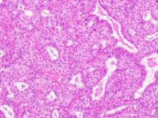
          Papillary neoplasms are a distinct assemblage of breast lesions whose main characteristic is the presence of fibrovascular cores which are surrounded by epithelial cells. Papillary lesions are of heterogenous nature, with similar clinical behavior and histomorphologic characteristics. Their biological patterns, however, can be quite different. According to the World Health Organization (WHO) (2019), breast tumors have been recently classified into five subdivisions of papillary neoplasms. They are namely: intraductal papilloma, papillary ductal carcinoma in situ, encapsulated papillary carcinoma (EPC), solid-papillary carcinoma and invasive papillary carcinoma. Despite the papillary architecture being easily recognized, histological variations are diagnostically challenging. The presence or absence of myoepithelial cells in the papillary cores can distinguish the malignant from the benign lesions respectively. EPC is a rare, histologically unique carcinoma type whose main characteristic is a thick fibrous capsule at the periphery and a prolific cell structure with fibrovascular stalk support. A characteristic feature is the absence of myoepithelial cells at the surrounding thick fibrous capsule. Usually, EPC maintains a slowly developing tumor despite the absence of myoepithelial cells. An EPC case presents diagnostic difficulties since it bears close resemblance to malignant and benign papillary breast lesions. Upon a clinical and radiological evaluation, EPC commonly appears as a benign lump. In mammography, the tumor is frequently found in a retroareolar position as a well-defined mass. On the other hand, in an ultrasound, the tumor will appear as a cystic lesion characterized by solid components. The clinical picture of EPC is usually an asymptomatic benign mass which at times can be felt through auto-palpation or screening mammography. A bloody nipple discharge is regarded as a common symptom. We report a case of an EPC of a 81-year-old woman who presented with a mass in the left breast.

          Related collections

          Most cited references22

          • Record: found
          • Abstract: found
          • Article: not found

          Intracystic papillary carcinoma: a review of 917 cases.

          Intracystic papillary carcinoma (IPC) is an uncommon breast neoplasm. To the authors' knowledge there are limited data regarding its epidemiology and only small studies focusing on outcomes. By using a large, population-based database, this study aimed to identify specific characteristics of patients with IPC, investigate its natural history, and determine its long-term prognosis. The California Cancer Registry (CCR), a population-based registry, was reviewed from the years 1988 through 2005. The data were analyzed with regard to patient sex, age at presentation, tumor stage, and overall survival. Cumulative relative actuarial survival was determined using a Berkson-Gage life table method. The CCR classifies IPC as either in situ (CIS) or invasive, as determined by the local pathologist. A total of 917 cases of IPC were identified. Approximately 47% of cases (n = 427) were CIS, whereas 53% of cases had invasion (n = 490). The majority of the invasive cases were localized at the time of diagnosis (89.6%; n = 439). At 10 years, patients with CIS and invasive disease had a similar relative cumulative survival (96.8% and 94.4%; P = .18). IPC is a rare disease. There is no apparently significant difference in the long-term survival of patients in the 2 histologically derived subgroups of IPC. There is an excellent prognosis for patients diagnosed with IPC regardless of whether the tumor is diagnosed as in situ or invasive. Clinicians should keep this in mind when planning surgical and adjuvant treatments. Sentinel lymph node biopsy may be a prudent way to evaluate axillary involvement in patients with IPC. (c) 2008 American Cancer Society.
            Bookmark
            • Record: found
            • Abstract: found
            • Article: not found

            Papillary lesions of the breast: MRI, ultrasound, and mammographic appearances.

            The purpose of this article is to describe the different imaging appearances of benign and malignant papillary lesions of the breast as well as to point out potential errors of interpretation that can lead to misdiagnosis. There is a wide spectrum of appearances of papillary lesions of the breast on MRI, ultrasound, and mammography. This variable appearance of papillary lesions makes differentiation of benign from malignant pathologies difficult on imaging, and tissue sampling is usually warranted.
              Bookmark
              • Record: found
              • Abstract: not found
              • Article: not found

              Papillary Lesions of the Breast: An Update

                Bookmark

                Author and article information

                Contributors
                Journal
                Front Surg
                Front Surg
                Front. Surg.
                Frontiers in Surgery
                Frontiers Media S.A.
                2296-875X
                04 February 2022
                2021
                : 8
                : 743881
                Affiliations
                Department of Obstetrics and Gynecology, Fribourg Cantonal Hospital Chemin des Pensionnats , Villars-sur-Glâne, Switzerland
                Author notes

                Edited by: Jean Marc bernard Ayoubi, Hôpital Foch, France

                Reviewed by: Manuel Maria Ianieri, Agostino Gemelli University Polyclinic (IRCCS), Italy; Ali Coskun, Izmir Bozyaka Training and Research Hospital, Turkey

                *Correspondence: Aikaterini Athanasiou katerinaioannathan@ 123456gmail.com

                This article was submitted to Obstetrics and Gynecology, a section of the journal Frontiers in Surgery

                Article
                10.3389/fsurg.2021.743881
                8854492
                35187043
                290a3d57-6736-44db-961c-c5c353582dff
                Copyright © 2022 Athanasiou, Khomsi, de Joliniere and Feki.

                This is an open-access article distributed under the terms of the Creative Commons Attribution License (CC BY). The use, distribution or reproduction in other forums is permitted, provided the original author(s) and the copyright owner(s) are credited and that the original publication in this journal is cited, in accordance with accepted academic practice. No use, distribution or reproduction is permitted which does not comply with these terms.

                History
                : 19 July 2021
                : 14 December 2021
                Page count
                Figures: 3, Tables: 0, Equations: 0, References: 22, Pages: 4, Words: 2907
                Categories
                Surgery
                Case Report

                breast cancer,encapsulated breast cancer,fibrovascular cores,sentinel lymph node,biopsy,receptors

                Comments

                Comment on this article