3
views
0
recommends
+1 Recommend
0 collections
    0
    shares
      • Record: found
      • Abstract: found
      • Article: found
      Is Open Access

      Local Progression Kinetics of Geographic Atrophy Depends Upon the Border Location

      research-article

      Read this article at

      Bookmark
          There is no author summary for this article yet. Authors can add summaries to their articles on ScienceOpen to make them more accessible to a non-specialist audience.

          Abstract

          Purpose

          To assess the influence of lesion morphology and location on geographic atrophy (GA) growth rate.

          Methods

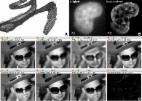
          We manually delineated GA on color fundus photographs of 237 eyes in the Age-Related Eye Disease Study. We calculated local border expansion rate (BER) as the linear distance that a point on the GA border traveled over 1 year based on a Euclidean distance map. Eye-specific BER was defined as the mean local BER of all points on the GA border in an eye. The percentage area affected by GA was defined as the GA area divided by the total retinal area in the region.

          Results

          GA enlarged 1.51 ± 1.96 mm 2 in area and 0.13 ± 0.11 mm in distance over 1 year. The GA area growth rate (mm 2/y) was associated with the baseline GA area ( P < 0.001), perimeter ( P < 0.001), lesion number ( P < 0.001), and circularity index ( P < 0.001); in contrast, eye-specific BER (mm/y) was not significantly associated with any of these factors. As the retinal eccentricity increased from 0 to 3.5 mm, the local BER increased from 0.10 to 0.24 mm/y ( P < 0.001); in contrast, the percentage of area affected by GA decreased from 49.3% to 2.3%.

          Conclusions

          Using distance-based measurements allows GA progression evaluation without significant confounding effects from baseline GA morphology. Local GA progression rates increased as a function of retinal eccentricity within the macula which is opposite of the trend for GA distribution, suggesting that GA initiation and enlargement may be mediated by different biological processes.

          Related collections

          Most cited references76

          • Record: found
          • Abstract: found
          • Article: found
          Is Open Access

          ImageJ2: ImageJ for the next generation of scientific image data

          Background ImageJ is an image analysis program extensively used in the biological sciences and beyond. Due to its ease of use, recordable macro language, and extensible plug-in architecture, ImageJ enjoys contributions from non-programmers, amateur programmers, and professional developers alike. Enabling such a diversity of contributors has resulted in a large community that spans the biological and physical sciences. However, a rapidly growing user base, diverging plugin suites, and technical limitations have revealed a clear need for a concerted software engineering effort to support emerging imaging paradigms, to ensure the software’s ability to handle the requirements of modern science. Results We rewrote the entire ImageJ codebase, engineering a redesigned plugin mechanism intended to facilitate extensibility at every level, with the goal of creating a more powerful tool that continues to serve the existing community while addressing a wider range of scientific requirements. This next-generation ImageJ, called “ImageJ2” in places where the distinction matters, provides a host of new functionality. It separates concerns, fully decoupling the data model from the user interface. It emphasizes integration with external applications to maximize interoperability. Its robust new plugin framework allows everything from image formats, to scripting languages, to visualization to be extended by the community. The redesigned data model supports arbitrarily large, N-dimensional datasets, which are increasingly common in modern image acquisition. Despite the scope of these changes, backwards compatibility is maintained such that this new functionality can be seamlessly integrated with the classic ImageJ interface, allowing users and developers to migrate to these new methods at their own pace. Conclusions Scientific imaging benefits from open-source programs that advance new method development and deployment to a diverse audience. ImageJ has continuously evolved with this idea in mind; however, new and emerging scientific requirements have posed corresponding challenges for ImageJ’s development. The described improvements provide a framework engineered for flexibility, intended to support these requirements as well as accommodate future needs. Future efforts will focus on implementing new algorithms in this framework and expanding collaborations with other popular scientific software suites. Electronic supplementary material The online version of this article (doi:10.1186/s12859-017-1934-z) contains supplementary material, which is available to authorized users.
            Bookmark
            • Record: found
            • Abstract: found
            • Article: not found
            Is Open Access

            Global prevalence of age-related macular degeneration and disease burden projection for 2020 and 2040: a systematic review and meta-analysis.

            Numerous population-based studies of age-related macular degeneration have been reported around the world, with the results of some studies suggesting racial or ethnic differences in disease prevalence. Integrating these resources to provide summarised data to establish worldwide prevalence and to project the number of people with age-related macular degeneration from 2020 to 2040 would be a useful guide for global strategies. We did a systematic literature review to identify all population-based studies of age-related macular degeneration published before May, 2013. Only studies using retinal photographs and standardised grading classifications (the Wisconsin age-related maculopathy grading system, the international classification for age-related macular degeneration, or the Rotterdam staging system) were included. Hierarchical Bayesian approaches were used to estimate the pooled prevalence, the 95% credible intervals (CrI), and to examine the difference in prevalence by ethnicity (European, African, Hispanic, Asian) and region (Africa, Asia, Europe, Latin America and the Caribbean, North America, and Oceania). UN World Population Prospects were used to project the number of people affected in 2014 and 2040. Bayes factor was calculated as a measure of statistical evidence, with a score above three indicating substantial evidence. Analysis of 129,664 individuals (aged 30-97 years), with 12,727 cases from 39 studies, showed the pooled prevalence (mapped to an age range of 45-85 years) of early, late, and any age-related macular degeneration to be 8.01% (95% CrI 3.98-15.49), 0.37% (0.18-0.77), and 8.69% (4.26-17.40), respectively. We found a higher prevalence of early and any age-related macular degeneration in Europeans than in Asians (early: 11.2% vs 6.8%, Bayes factor 3.9; any: 12.3% vs 7.4%, Bayes factor 4.3), and early, late, and any age-related macular degeneration to be more prevalent in Europeans than in Africans (early: 11.2% vs 7.1%, Bayes factor 12.2; late: 0.5% vs 0.3%, 3.7; any: 12.3% vs 7.5%, 31.3). There was no difference in prevalence between Asians and Africans (all Bayes factors <1). Europeans had a higher prevalence of geographic atrophy subtype (1.11%, 95% CrI 0.53-2.08) than Africans (0.14%, 0.04-0.45), Asians (0.21%, 0.04-0.87), and Hispanics (0.16%, 0.05-0.46). Between geographical regions, cases of early and any age-related macular degeneration were less prevalent in Asia than in Europe and North America (early: 6.3% vs 14.3% and 12.8% [Bayes factor 2.3 and 7.6]; any: 6.9% vs 18.3% and 14.3% [3.0 and 3.8]). No significant gender effect was noted in prevalence (Bayes factor <1.0). The projected number of people with age-related macular degeneration in 2020 is 196 million (95% CrI 140-261), increasing to 288 million in 2040 (205-399). These estimates indicate the substantial global burden of age-related macular degeneration. Summarised data provide information for understanding the effect of the condition and provide data towards designing eye-care strategies and health services around the world. National Medical Research Council, Singapore. Copyright © 2014 Wong et al. Open Access article distributed under the terms of CC BY-NC-ND. Published by .. All rights reserved.
              Bookmark
              • Record: found
              • Abstract: found
              • Article: found

              A large genome-wide association study of age-related macular degeneration highlights contributions of rare and common variants

              Advanced age-related macular degeneration (AMD) is the leading cause of blindness in the elderly with limited therapeutic options. Here, we report on a study of >12 million variants including 163,714 directly genotyped, most rare, protein-altering variant. Analyzing 16,144 patients and 17,832 controls, we identify 52 independently associated common and rare variants (P < 5×10⁻⁸) distributed across 34 loci. While wet and dry AMD subtypes exhibit predominantly shared genetics, we identify the first signal specific to wet AMD, near MMP9 (difference-P = 4.1×10⁻¹⁰). Very rare coding variants (frequency < 0.1%) in CFH, CFI, and TIMP3 suggest causal roles for these genes, as does a splice variant in SLC16A8. Our results support the hypothesis that rare coding variants can pinpoint causal genes within known genetic loci and illustrate that applying the approach systematically to detect new loci requires extremely large sample sizes.
                Bookmark

                Author and article information

                Journal
                Invest Ophthalmol Vis Sci
                Invest Ophthalmol Vis Sci
                iovs
                IOVS
                Investigative Ophthalmology & Visual Science
                The Association for Research in Vision and Ophthalmology
                0146-0404
                1552-5783
                28 October 2021
                October 2021
                : 62
                : 13
                : 28
                Affiliations
                [1 ]Department of Ophthalmology, University of California, San Francisco, San Francisco, California, United States
                [2 ]Institute of Cardiovascular Diseases, Gladstone Institute, San Francisco, California, United States
                [3 ]Byers Eye Institute, Department of Ophthalmology, Stanford University School of Medicine, Palo Alto, California, United States
                [4 ]Department of Ophthalmology, New York Eye and Ear Infirmary of Mount Sinai, New York, New York, United States
                [5 ]Department of Ophthalmology and Visual Science, Kellogg Eye Center, University of Michigan Medical School, Ann Arbor, Michigan, United States
                [6 ]Department of Ophthalmology and Visual Science, Yale School of Medicine, New Haven, Connecticut, United States
                Author notes
                Correspondence: Lucian V. Del Priore, Department of Ophthalmology and Visual Science, Yale School of Medicine, 40 Temple Street, Suite 1B, New Haven, CT 06510, USA; lucian.delpriore@ 123456yale.edu .
                Article
                IOVS-21-32715
                10.1167/iovs.62.13.28
                8558522
                34709347
                28f843d7-94d7-432a-8fee-9fcfa2e77207
                Copyright 2021 The Authors

                This work is licensed under a Creative Commons Attribution 4.0 International License.

                History
                : 06 October 2021
                : 04 April 2021
                Page count
                Pages: 11
                Categories
                Retina
                Retina

                age-related macular degeneration,geographic atrophy,natural history,color fundus photography

                Comments

                Comment on this article