0
views
0
recommends
+1 Recommend
0 collections
    0
    shares
      • Record: found
      • Abstract: found
      • Article: found
      Is Open Access

      Convolution neural network for the diagnosis of wireless capsule endoscopy: a systematic review and meta-analysis

      review-article

      Read this article at

      Bookmark
          There is no author summary for this article yet. Authors can add summaries to their articles on ScienceOpen to make them more accessible to a non-specialist audience.

          Abstract

          Background

          Wireless capsule endoscopy (WCE) is considered to be a powerful instrument for the diagnosis of intestine diseases. Convolution neural network (CNN) is a type of artificial intelligence that has the potential to assist the detection of WCE images. We aimed to perform a systematic review of the current research progress to the CNN application in WCE.

          Methods

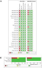
          A search in PubMed, SinoMed, and Web of Science was conducted to collect all original publications about CNN implementation in WCE. Assessment of the risk of bias was performed by Quality Assessment of Diagnostic Accuracy Studies-2 risk list. Pooled sensitivity and specificity were calculated by an exact binominal rendition of the bivariate mixed-effects regression model. I 2 was used for the evaluation of heterogeneity.

          Results

          16 articles with 23 independent studies were included. CNN application to WCE was divided into detection on erosion/ulcer, gastrointestinal bleeding (GI bleeding), and polyps/cancer. The pooled sensitivity of CNN for erosion/ulcer is 0.96 [95% CI 0.91, 0.98], for GI bleeding is 0.97 (95% CI 0.93–0.99), and for polyps/cancer is 0.97 (95% CI 0.82–0.99). The corresponding specificity of CNN for erosion/ulcer is 0.97 (95% CI 0.93–0.99), for GI bleeding is 1.00 (95% CI 0.99–1.00), and for polyps/cancer is 0.98 (95% CI 0.92–0.99).

          Conclusion

          Based on our meta-analysis, CNN-dependent diagnosis of erosion/ulcer, GI bleeding, and polyps/cancer approached a high-level performance because of its high sensitivity and specificity. Therefore, future perspective, CNN has the potential to become an important assistant for the diagnosis of WCE.

          Supplementary Information

          The online version contains supplementary material available at 10.1007/s00464-021-08689-3.

          Related collections

          Most cited references51

          • Record: found
          • Abstract: not found
          • Article: not found

          GRADE: an emerging consensus on rating quality of evidence and strength of recommendations.

            Bookmark
            • Record: found
            • Abstract: found
            • Article: found
            Is Open Access

            PRISMA 2020 explanation and elaboration: updated guidance and exemplars for reporting systematic reviews

            The methods and results of systematic reviews should be reported in sufficient detail to allow users to assess the trustworthiness and applicability of the review findings. The Preferred Reporting Items for Systematic reviews and Meta-Analyses (PRISMA) statement was developed to facilitate transparent and complete reporting of systematic reviews and has been updated (to PRISMA 2020) to reflect recent advances in systematic review methodology and terminology. Here, we present the explanation and elaboration paper for PRISMA 2020, where we explain why reporting of each item is recommended, present bullet points that detail the reporting recommendations, and present examples from published reviews. We hope that changes to the content and structure of PRISMA 2020 will facilitate uptake of the guideline and lead to more transparent, complete, and accurate reporting of systematic reviews.
              Bookmark
              • Record: found
              • Abstract: found
              • Article: not found

              Artificial intelligence in radiology

              Artificial intelligence (AI) algorithms, particularly deep learning, have demonstrated remarkable progress in image-recognition tasks. Methods ranging from convolutional neural networks to variational autoencoders have found myriad applications in the medical image analysis field, propelling it forward at a rapid pace. Historically, in radiology practice, trained physicians visually assessed medical images for the detection, characterization and monitoring of diseases. AI methods excel at automatically recognizing complex patterns in imaging data and providing quantitative, rather than qualitative, assessments of radiographic characteristics. In this O pinion article, we establish a general understanding of AI methods, particularly those pertaining to image-based tasks. We explore how these methods could impact multiple facets of radiology, with a general focus on applications in oncology, and demonstrate ways in which these methods are advancing the field. Finally, we discuss the challenges facing clinical implementation and provide our perspective on how the domain could be advanced.
                Bookmark

                Author and article information

                Contributors
                liqingyuan09@smu.edu.cn
                Journal
                Surg Endosc
                Surg Endosc
                Surgical Endoscopy
                Springer US (New York )
                0930-2794
                1432-2218
                23 August 2021
                23 August 2021
                2022
                : 36
                : 1
                : 16-31
                Affiliations
                [1 ]GRID grid.284723.8, ISNI 0000 0000 8877 7471, Nanfang Hospital (The First School of Clinical Medicine), , Southern Medical University, ; Guangzhou, Guangdong China
                [2 ]Guangzhou SiDe MedTech Co.,Ltd, Guangzhou, Guangdong China
                [3 ]GRID grid.284723.8, ISNI 0000 0000 8877 7471, Guangdong Provincial Key Laboratory of Gastroenterology, Department of Gastroenterology, Nanfang Hospital, , Southern Medical University, ; No. 1838, Guangzhou Avenue North, Guangzhou, Guangdong China
                [4 ]GRID grid.284723.8, ISNI 0000 0000 8877 7471, State Key Laboratory of Organ Failure Research, Guangdong Provincial Key Laboratory of Viral Hepatitis Research, Department of Hepatology Unit and Infectious Diseases, Nanfang Hospital, , Southern Medical University, ; Guangzhou, Guangdong China
                Author information
                http://orcid.org/0000-0002-5601-8467
                Article
                8689
                10.1007/s00464-021-08689-3
                8741689
                34426876
                28cbba63-d7bc-4029-87ad-a0e932462f46
                © The Author(s) 2021

                Open AccessThis article is licensed under a Creative Commons Attribution 4.0 International License, which permits use, sharing, adaptation, distribution and reproduction in any medium or format, as long as you give appropriate credit to the original author(s) and the source, provide a link to the Creative Commons licence, and indicate if changes were made. The images or other third party material in this article are included in the article's Creative Commons licence, unless indicated otherwise in a credit line to the material. If material is not included in the article's Creative Commons licence and your intended use is not permitted by statutory regulation or exceeds the permitted use, you will need to obtain permission directly from the copyright holder. To view a copy of this licence, visit http://creativecommons.org/licenses/by/4.0/.

                History
                : 5 May 2021
                : 7 August 2021
                Funding
                Funded by: guangdong basic and applied basic research fund
                Award ID: 2021A1515010992
                Funded by: National Natural Science Funds
                Award ID: 12026605
                Funded by: Guangdong Basic and Applied Basic Research Fund
                Award ID: 2020A1515110916
                Funded by: Guangdong Medical Science and Technology Research Fund Project
                Award ID: A2020143
                Funded by: 2020 Southern Medical University Innovation and Entrepreneurship Training Program
                Award ID: 202012121035X
                Funded by: Foundation for the President of Nanfang Hospital of Southern Medical University
                Award ID: 2018C027
                Funded by: Guangdong Science and Technology Plan Project
                Award ID: 2017B020209003
                Categories
                Review Article
                Custom metadata
                © Springer Science+Business Media, LLC, part of Springer Nature 2022

                Surgery
                deep learning,convolutional neural network,capsule endoscopy
                Surgery
                deep learning, convolutional neural network, capsule endoscopy

                Comments

                Comment on this article