39
views
0
recommends
+1 Recommend
0 collections
    0
    shares
      • Record: found
      • Abstract: found
      • Article: not found

      Deciphering the rules governing assembly order of mammalian septin complexes

      research-article

      Read this article at

      ScienceOpenPublisherPMC
      Bookmark
          There is no author summary for this article yet. Authors can add summaries to their articles on ScienceOpen to make them more accessible to a non-specialist audience.

          Abstract

          Vertebrates express 9–17 septin family members known to oligomerize into diverse structures, but their native assembly states have remained elusive. The results presented suggest a generic model for how the temporal order of septin assembly directs the subunit arrangement within distinct pools of six- to eight-subunit core heteromers.

          Abstract

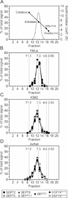
          Septins are conserved GTP-binding proteins that assemble into lateral diffusion barriers and molecular scaffolds. Vertebrate genomes contain 9–17 septin genes that encode both ubiquitous and tissue-specific septins. Expressed septins may assemble in various combinations through both heterotypic and homotypic G-domain interactions. However, little is known regarding assembly states of mammalian septins and mechanisms directing ordered assembly of individual septins into heteromeric units, which is the focus of this study. Our analysis of the septin system in cells lacking or overexpressing selected septins reveals interdependencies coinciding with previously described homology subgroups. Hydrodynamic and single-particle data show that individual septins exist solely in the context of stable six- to eight-subunit core heteromers, all of which contain SEPT2 and SEPT6 subgroup members and SEPT7, while heteromers comprising more than six subunits also contain SEPT9. The combined data suggest a generic model for how the temporal order of septin assembly is homology subgroup-directed, which in turn determines the subunit arrangement of native heteromers. Because mammalian cells normally express multiple members and/or isoforms of some septin subgroups, our data also suggest that only a minor fraction of native heteromers are arranged as perfect palindromes.

          Related collections

          Most cited references25

          • Record: found
          • Abstract: found
          • Article: not found

          A septin diffusion barrier at the base of the primary cilium maintains ciliary membrane protein distribution.

          In animal cells, the primary cilium transduces extracellular signals through signaling receptors localized in the ciliary membrane, but how these ciliary membrane proteins are retained in the cilium is unknown. We found that ciliary membrane proteins were highly mobile, but their diffusion was impeded at the base of the cilium by a diffusion barrier. Septin 2 (SEPT2), a member of the septin family of guanosine triphosphatases that form a diffusion barrier in budding yeast, localized at the base of the ciliary membrane. SEPT2 depletion resulted in loss of ciliary membrane protein localization and Sonic hedgehog signal transduction, and inhibited ciliogenesis. Thus, SEPT2 is part of a diffusion barrier at the base of the ciliary membrane and is essential for retaining receptor-signaling pathways in the primary cilium.
            Bookmark
            • Record: found
            • Abstract: found
            • Article: not found

            Self- and actin-templated assembly of Mammalian septins.

            Septins are polymerizing GTPases required for cytokinesis and cortical organization. The principles by which they are targeted to, and assemble at, specific cell regions are unknown. We show that septins in mammalian cells switch between a linear organization along actin bundles and cytoplasmic rings, approximately 0.6 microm in diameter. A recombinant septin complex self-assembles into rings resembling those in cells. Linear organization along actin bundles was reconstituted by adding an adaptor protein, anillin. Perturbation of septin organization in cells by expression of a septin-interacting fragment of anillin or by septin depletion via siRNA causes loss of actin bundles. We conclude that septins alone self-assemble into rings, that adaptor proteins recruit septins to actin bundles, and that septins help organize these bundles.
              Bookmark
              • Record: found
              • Abstract: found
              • Article: not found

              The septin family of GTPases: architecture and dynamics.

              Septins comprise a conserved family of proteins that are found primarily in fungi and animals. These GTP-binding proteins have several roles during cell division, cytoskeletal organization and membrane-remodelling events. One factor that is crucial for their functions is the ordered assembly of individual septins into oligomeric core complexes that, in turn, form higher-order structures such as filaments, rings and gauzes. The molecular details of these interactions and the mechanism by which septin-complex assembly is regulated have remained elusive. Recently, the first detailed structural views of the septin core have emerged, and these, along with studies of septin dynamics in vivo, have provided new insight into septin-complex assembly and septin function in vivo.
                Bookmark

                Author and article information

                Contributors
                Role: Monitoring Editor
                Journal
                Mol Biol Cell
                molbiolcell
                mbc
                Mol. Bio. Cell
                Molecular Biology of the Cell
                The American Society for Cell Biology
                1059-1524
                1939-4586
                01 September 2011
                : 22
                : 17
                : 3152-3164
                Affiliations
                [1] aDepartment of Molecular Biology, Umeå University, SE-901 87 Umeå, Sweden
                [2] bDepartment of Cell and Molecular Biology, Karolinska Institute, S-171 77 Stockholm, Sweden
                University of California, Santa Cruz
                Author notes
                *Address correspondence to: Mikael E. Sellin ( mikael.sellin@ 123456molbiol.umu.se ).
                Article
                E11-03-0253
                10.1091/mbc.E11-03-0253
                3164462
                21737677
                27c50b07-775e-400a-a208-948224c6c062
                © 2011 Sellin et al. This article is distributed by The American Society for Cell Biology under license from the author(s). Two months after publication it is available to the public under an Attribution–Noncommercial–Share Alike 3.0 Unported Creative Commons License ( http://creativecommons.org/licenses/by-nc-sa/3.0).

                “ASCB®,” “The American Society for Cell Biology®,” and “Molecular Biology of the Cell®” are registered trademarks of The American Society of Cell Biology.

                History
                : 25 March 2011
                : 14 June 2011
                : 28 June 2011
                Categories
                Articles
                Cytoskeleton

                Molecular biology
                Molecular biology

                Comments

                Comment on this article