35
views
0
recommends
+1 Recommend
0 collections
    8
    shares
      • Record: found
      • Abstract: found
      • Article: found
      Is Open Access

      Antiapoptotic Effects of EGb 761

      review-article

      Read this article at

      Bookmark
          There is no author summary for this article yet. Authors can add summaries to their articles on ScienceOpen to make them more accessible to a non-specialist audience.

          Abstract

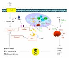
          Ginkgo biloba extracts have long been used in Chinese traditional medicine for hundreds of years. The most significant extract obtained from Ginkgo biloba leaves has been EGb 761, a widely used phytopharmaceutical product in Europe. EGb 761 is a well-defined mixture of active compounds, which contains two main active substances: flavonoid glycosides (24–26%) and terpene lactones (6–8%). These compounds have shown antiapoptotic effects through the protection of mitochondrial membrane integrity, inhibition of mitochondrial cytochrome c release, enhancement of antiapoptotic protein transcription, and reduction of caspase transcription and DNA fragmentation. Other effects include the reduction of oxidative stress (which has been related to the occurrence of vascular, degenerative, and proliferative diseases), coupled to strong induction of phase II-detoxifying and cellular defense enzymes by Nrf2/ARE activation, in addition to the modulation of transcription factors, such as CREB, HIF-1 α , NF- κ B, AP-1, and p53, involved in the apoptosis process. This work reviews experimental results about the antiapoptotic effects induced by the standardized extract of Ginkgo biloba leaves (EGb 761).

          Related collections

          Most cited references171

          • Record: found
          • Abstract: found
          • Article: not found

          Caspases: the executioners of apoptosis.

          Apoptosis is a major form of cell death, characterized initially by a series of stereotypic morphological changes. In the nematode Caenorhabditis elegans, the gene ced-3 encodes a protein required for developmental cell death. Since the recognition that CED-3 has sequence identity with the mammalian cysteine protease interleukin-1 beta-converting enzyme (ICE), a family of at least 10 related cysteine proteases has been identified. These proteins are characterized by almost absolute specificity for aspartic acid in the P1 position. All the caspases (ICE-like proteases) contain a conserved QACXG (where X is R, Q or G) pentapeptide active-site motif. Capases are synthesized as inactive proenzymes comprising an N-terminal peptide (prodomain) together with one large and one small subunit. The crystal structures of both caspase-1 and caspase-3 show that the active enzyme is a heterotetramer, containing two small and two large subunits. Activation of caspases during apoptosis results in the cleavage of critical cellular substrates, including poly(ADP-ribose) polymerase and lamins, so precipitating the dramatic morphological changes of apoptosis. Apoptosis induced by CD95 (Fas/APO-1) and tumour necrosis factor activates caspase-8 (MACH/FLICE/Mch5), which contains an N-terminus with FADD (Fas-associating protein with death domain)-like death effector domains, so providing a direct link between cell death receptors and the caspases. The importance of caspase prodomains in the regulation of apoptosis is further highlighted by the recognition of adapter molecules, such as RAIDD [receptor-interacting protein (RIP)-associated ICH-1/CED-3-homologous protein with a death domain]/CRADD (caspase and RIP adapter with death domain), which binds to the prodomain of caspase-2 and recruits it to the signalling complex. Cells undergoing apoptosis following triggering of death receptors execute the death programme by activating a hierarchy of caspases, with caspase-8 and possibly caspase-10 being at or near the apex of this apoptotic cascade.
            Bookmark
            • Record: found
            • Abstract: found
            • Article: not found

            Reactive oxygen species promote TNFalpha-induced death and sustained JNK activation by inhibiting MAP kinase phosphatases.

            TNFalpha is a pleiotropic cytokine that induces either cell proliferation or cell death. Inhibition of NF-kappaB activation increases susceptibility to TNFalpha-induced death, concurrent with sustained JNK activation, an important contributor to the death response. Sustained JNK activation in NF-kappaB-deficient cells was suggested to depend on reactive oxygen species (ROS), but how ROS affect JNK activation was unclear. We now show that TNFalpha-induced ROS, whose accumulation is suppressed by mitochondrial superoxide dismutase, cause oxidation and inhibition of JNK-inactivating phosphatases by converting their catalytic cysteine to sulfenic acid. This results in sustained JNK activation, which is required for cytochrome c release and caspase 3 cleavage, as well as necrotic cell death. Treatment of cells or experimental animals with an antioxidant prevents H(2)O(2) accumulation, JNK phosphatase oxidation, sustained JNK activity, and both forms of cell death. Antioxidant treatment also prevents TNFalpha-mediated fulminant liver failure without affecting liver regeneration.
              Bookmark
              • Record: found
              • Abstract: found
              • Article: not found

              Apaf-1, a human protein homologous to C. elegans CED-4, participates in cytochrome c-dependent activation of caspase-3.

              We report here the purification and cDNA cloning of Apaf-1, a novel 130 kd protein from HeLa cell cytosol that participates in the cytochrome c-dependent activation of caspase-3. The NH2-terminal 85 amino acids of Apaf-1 show 21% identity and 53% similarity to the NH2-terminal prodomain of the Caenorhabditis elegans caspase, CED-3. This is followed by 320 amino acids that show 22% identity and 48% similarity to CED-4, a protein that is believed to initiate apoptosis in C. elegans. The COOH-terminal region of Apaf-1 comprises multiple WD repeats, which are proposed to mediate protein-protein interactions. Cytochrome c binds to Apaf-1, an event that may trigger the activation of caspase-3, leading to apoptosis.
                Bookmark

                Author and article information

                Journal
                Evid Based Complement Alternat Med
                Evid Based Complement Alternat Med
                ECAM
                Evidence-based Complementary and Alternative Medicine : eCAM
                Hindawi Publishing Corporation
                1741-427X
                1741-4288
                2013
                29 July 2013
                29 July 2013
                : 2013
                : 495703
                Affiliations
                1Laboratorio de Neurobiología Molecular y Celular, INNN-UNAM, Instituto Nacional de Neurología y Neurocirugía “Manuel Velasco Suárez”, 14269 México, DF, Mexico
                2Departamento de Biología, Facultad de Química, Universidad Nacional Autónoma de México, 04510 México, DF, Mexico
                3Laboratorio Analítico de Compuestos del Tabaco, Instituto Nacional de Salud Pública, 014000 México, DF, Mexico
                4Departamento de Salud Bucal, Centro Nacional de Programas Preventivo y Control de Enfermedades, 11800 México, DF, Mexico
                5Área de Neurociencias, Departamento de Biología de la Reproducción, Universidad Autónoma Metropolitana-Iztapalapa, 09340 México, DF, Mexico
                6Departamento de Neuroinmunología, Instituto Nacional de Neurología y Neurocirugía “Manuel Velasco Suárez”, 14269 México, DF, Mexico
                Author notes
                *Cristina Trejo-Solís: trejosolis@ 123456yahoo.com.mx

                Academic Editor: Alfredo Vannacci

                Article
                10.1155/2013/495703
                3745884
                23983787
                276b3172-6845-4345-9b37-a9801f22523e
                Copyright © 2013 Norma Serrano-García et al.

                This is an open access article distributed under the Creative Commons Attribution License, which permits unrestricted use, distribution, and reproduction in any medium, provided the original work is properly cited.

                History
                : 19 February 2013
                : 19 June 2013
                : 27 June 2013
                Categories
                Review Article

                Complementary & Alternative medicine
                Complementary & Alternative medicine

                Comments

                Comment on this article