13
views
0
recommends
+1 Recommend
0 collections
    0
    shares
      • Record: found
      • Abstract: found
      • Article: found
      Is Open Access

      Intraosseous angiosarcoma with secondary aneurysmal bone cysts presenting as an elusive diagnostic challenge

      research-article

      Read this article at

      Bookmark
          There is no author summary for this article yet. Authors can add summaries to their articles on ScienceOpen to make them more accessible to a non-specialist audience.

          Abstract

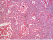
          Angiosarcoma of bone is an exceedingly rare primary bone malignancy that can present as an aggressive osteolytic lesion. Histological diagnosis can be extremely challenging, as the pathological features often resemble that of aneurysmal bone cysts. We report an interesting and peculiar case of an intraosseous angiosarcoma that presented as a diagnostic dilemma and discuss the relevant radiological and pathologic findings.

          Related collections

          Most cited references24

          • Record: found
          • Abstract: found
          • Article: not found

          Aneurysmal bone cyst: a review of 150 patients.

          We have reviewed a series of 150 aneurysmal bone cysts treated over the last 20 years. The lesions were principally located in the tibia, femur, pelvis, humerus, and spine and, in most cases, presented the imaging appearance originally described by Jaffe and Lichtenstein as a blowout with thin cortices. Only one of the patients was believed to have an osteoblastoma of the spine with secondary development of an aneurysmal bone cyst, and none of the patients developed additional lesions. The patients were treated primarily with curettage and implantation of allograft chips or polymethylmethacrylate, but some patients were treated with insertion of autografts or allografts. The local recurrence rate was 20%, which is consistent with that reported by other centers. Aneurysmal bone cysts are enigmatic lesions of unknown cause and presentation and are difficult to distinguish from other lesions. Overall, the treatment is satisfactory, but it is possible that newer approaches, such as improved magnetic resonance imaging studies, may help diagnose the lesions and allow the physicians to plan for more effective treatment protocols.
            Bookmark
            • Record: found
            • Abstract: found
            • Article: not found

            Bone cancers.

            Frequency distribution data for primary bone sarcomas have long been used to provide clues to the diagnosis of bone cancers after their identification in radiographs. Age and skeletal site are often helpful, in addition to specific radiographic features, in narrowing down the probable histologic categories of bone neoplasms before biopsy. Data on 2627 histologically confirmed primary malignant tumors of bone, as collected by the SEER program during the period 1973-1987, were analyzed by age, sex, race, and anatomical site. Osteosarcoma was the most frequently diagnosed primary sarcoma of bone (35.1%), followed by chondrosarcoma (25.8%), Ewing's sarcoma (16.0%), chordoma (8.4%), and malignant fibrous histiocytoma, including fibrosarcoma (5.6%). The most frequently diagnosed sarcoma of bone in persons younger than age 20 was osteosarcoma, followed by Ewing's sarcoma. Chondrosarcoma was the most frequently diagnosed bone tumor in the population older than age 50. The overall 5-year relative survival rates were as follows: osteosarcoma, 41.0%; chondrosarcoma, 72.7%; Ewing's sarcoma, 41.2%; chordoma, 63.8%; and malignant fibrous histiocytoma, 42.9%. There was an improvement in the survival rates during the period 1973-1987 for patients with chordoma and for white female patients with Ewing's sarcoma. Ewing's sarcoma and chordoma occurred almost exclusively in the white population. SEER data provide a unique opportunity to evaluate the incidence and survival rates of bone sarcomas, which are uncommon but highly lethal tumors. The findings from this analysis provide information useful in the diagnosis of these cancers.
              Bookmark
              • Record: found
              • Abstract: found
              • Article: not found

              A 14-year retrospective review of angiosarcoma: clinical characteristics, prognostic factors, and treatment outcomes with surgery and chemotherapy.

              Angiosarcoma is a rare vascular malignancy, and there are few published data to guide chemotherapy treatment decisions. We present a retrospective analysis of angiosarcoma encompassing all anatomic sites of disease presenting to a single institution over a 14-year period. Characteristics at presentation and prognostic factors are reviewed. For patients with unresectable disease, progression-free survival with various chemotherapy regimens is described. Pathological confirmation of all cases was performed before they were included in this analysis. One hundred twenty-five patients with angiosarcoma were seen and treated between January 1, 1990 and December 31, 2003. Angiosarcoma showed marked variation by anatomic site regarding gender ratio, median age at diagnosis, overall survival, and response to chemotherapy. Overall 5-year survival was 31% for angiosarcoma. Superficial depth and negative microscopic surgical margins correlated with longer overall survival, but tumor size did not reach significance as a prognostic factor. For unresectable angiosarcoma, doxorubicin based regimens yielded progression-free survival of 3.7-5.4 months. Paclitaxel achieved a progression-free survival of 6.8 months for scalp angiosarcoma and 2.8 months for sites below the clavicle. Angiosarcoma is an aggressive malignancy characterized by biologic heterogeneity at different anatomic sites and relative sensitivity to paclitaxel and doxorubicin.
                Bookmark

                Author and article information

                Journal
                Int Semin Surg Oncol
                International seminars in surgical oncology : ISSO
                BioMed Central
                1477-7800
                2008
                20 May 2008
                : 5
                : 10
                Affiliations
                [1 ]Department of Orthopaedics, St. Vincent's Hospital, Melbourne, Australia
                [2 ]Bone and Soft Tissue Sarcoma Service, Peter MacCallum Cancer Centre, Melbourne, Australia
                [3 ]Department of Anatomical Pathology, St Vincent's Hospital, Melbourne, Australia
                [4 ]Department of Radiology, St. Vincent's Hospital, Melbourne, Australia
                Article
                1477-7800-5-10
                10.1186/1477-7800-5-10
                2413251
                18492283
                2760750e-262b-47d9-8575-4bd104367465
                Copyright © 2008 Tse et al; licensee BioMed Central Ltd.

                This is an Open Access article distributed under the terms of the Creative Commons Attribution License ( http://creativecommons.org/licenses/by/2.0), which permits unrestricted use, distribution, and reproduction in any medium, provided the original work is properly cited.

                History
                : 12 December 2007
                : 20 May 2008
                Categories
                Case Report

                Oncology & Radiotherapy
                Oncology & Radiotherapy

                Comments

                Comment on this article