49
views
0
recommends
+1 Recommend
0 collections
    0
    shares
      • Record: found
      • Abstract: found
      • Article: found
      Is Open Access

      Crystal structure of human polynucleotide phosphorylase: insights into its domain function in RNA binding and degradation

      research-article

      Read this article at

      Bookmark
          There is no author summary for this article yet. Authors can add summaries to their articles on ScienceOpen to make them more accessible to a non-specialist audience.

          Abstract

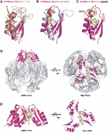
          Human polynucleotide phosphorylase (hPNPase) is a 3′-to-5′ exoribonuclease that degrades specific mRNA and miRNA, and imports RNA into mitochondria, and thus regulates diverse physiological processes, including cellular senescence and homeostasis. However, the RNA-processing mechanism by hPNPase, particularly how RNA is bound via its various domains, remains obscure. Here, we report the crystal structure of an S1 domain-truncated hPNPase at a resolution of 2.1 Å. The trimeric hPNPase has a hexameric ring-like structure formed by six RNase PH domains, capped with a trimeric KH pore. Our biochemical and mutagenesis studies suggest that the S1 domain is not critical for RNA binding, and conversely, that the conserved GXXG motif in the KH domain directly participates in RNA binding in hPNPase. Our studies thus provide structural and functional insights into hPNPase, which uses a KH pore to trap a long RNA 3′ tail that is further delivered into an RNase PH channel for the degradation process. Structural RNA with short 3′ tails are, on the other hand, transported but not digested by hPNPase.

          Related collections

          Most cited references51

          • Record: found
          • Abstract: found
          • Article: not found

          RNA-quality control by the exosome.

          The exosome complex of 3'-->5' exonucleases is an important component of the RNA-processing machinery in eukaryotes. This complex functions in the accurate processing of nuclear RNA precursors and in the degradation of RNAs in both the nucleus and the cytoplasm. However, it has been unclear how different classes of substrate are distinguished from one another. Recent studies now provide insights into the regulation and structure of the exosome, and they reveal striking similarities between the process of RNA degradation in bacteria and eukaryotes.
            Bookmark
            • Record: found
            • Abstract: found
            • Article: not found

            Structure and function of KH domains.

            The hnRNP K homology (KH) domain was first identified in the protein human heterogeneous nuclear ribonucleoprotein K (hnRNP K) 14 years ago. Since then, KH domains have been identified as nucleic acid recognition motifs in proteins that perform a wide range of cellular functions. KH domains bind RNA or ssDNA, and are found in proteins associated with transcriptional and translational regulation, along with other cellular processes. Several diseases, e.g. fragile X mental retardation syndrome and paraneoplastic disease, are associated with the loss of function of a particular KH domain. Here we discuss the progress made towards understanding both general and specific features of the molecular recognition of nucleic acids by KH domains. The typical binding surface of KH domains is a cleft that is versatile but that can typically accommodate only four unpaired bases. Van der Waals forces and hydrophobic interactions and, to a lesser extent, electrostatic interactions, contribute to the nucleic acid binding affinity. 'Augmented' KH domains or multiple copies of KH domains within a protein are two strategies that are used to achieve greater affinity and specificity of nucleic acid binding. Isolated KH domains have been seen to crystallize as monomers, dimers and tetramers, but no published data support the formation of noncovalent higher-order oligomers by KH domains in solution. Much attention has been given in the literature to a conserved hydrophobic residue (typically Ile or Leu) that is present in most KH domains. The interest derives from the observation that an individual with this Ile mutated to Asn, in the KH2 domain of fragile X mental retardation protein, exhibits a particularly severe form of the syndrome. The structural effects of this mutation in the fragile X mental retardation protein KH2 domain have recently been reported. We discuss the use of analogous point mutations at this position in other KH domains to dissect both structure and function.
              Bookmark
              • Record: found
              • Abstract: found
              • Article: not found

              Reconstitution, activities, and structure of the eukaryotic RNA exosome.

              The RNA exosome is a multisubunit 3' to 5' exoribonuclease complex that participates in degradation and processing of cellular RNA. To determine the activities and structure of the eukaryotic exosome, we report the reconstitution of 9-subunit exosomes from yeast and human and reconstitution of 10- and 11-subunit exosomes from yeast. Comparative biochemical analysis between purified subunits and reconstituted exosomes using AU-rich, polyadenylated (poly[A]), generic, and structured RNA substrates reveals processive phosphorolytic activities for human Rrp41/Rrp45 and the 9-subunit human exosome, processive hydrolytic activities for yeast Rrp44 and the yeast 10-subunit exosome, distributive hydrolytic activities for Rrp6, and processive and distributive hydrolytic activities for the yeast 11-subunit exosome. To elucidate the architecture of a eukaryotic exosome, its conserved surfaces, and the structural basis for RNA decay, we report the X-ray structure determination for the 286 kDa nine-subunit human exosome at 3.35 A.
                Bookmark

                Author and article information

                Journal
                Nucleic Acids Res
                Nucleic Acids Res
                nar
                nar
                Nucleic Acids Research
                Oxford University Press
                0305-1048
                1362-4962
                May 2012
                May 2012
                30 December 2011
                30 December 2011
                : 40
                : 9
                : 4146-4157
                Affiliations
                1Institute of Bioinformatics and Structural Biology, National Tsing Hua University, Hsinchu 30013, 2Institute of Molecular Biology, Academia Sinica, Taipei 11529 and 3Graduate Institute of Biochemistry and Molecular Biology, National Taiwan University, Taipei 10051, Taiwan, R.O.C.
                Author notes
                *To whom correspondence should be addressed. Tel: +886 2 27884151; Fax: +886 2 27826085; Email: hanna@ 123456sinica.edu.tw
                Article
                gkr1281
                10.1093/nar/gkr1281
                3351181
                22210891
                27101d38-eb24-4535-a659-d34baca04661
                © The Author(s) 2011. Published by Oxford University Press.

                This is an Open Access article distributed under the terms of the Creative Commons Attribution Non-Commercial License ( http://creativecommons.org/licenses/by-nc/3.0), which permits unrestricted non-commercial use, distribution, and reproduction in any medium, provided the original work is properly cited.

                History
                : 4 November 2011
                : 8 December 2011
                : 9 December 2011
                Page count
                Pages: 12
                Categories
                Structural Biology

                Genetics
                Genetics

                Comments

                Comment on this article