22
views
0
recommends
+1 Recommend
0 collections
    0
    shares
      • Record: found
      • Abstract: found
      • Article: found
      Is Open Access

      CSF A β42/A β40 and A β42/A β38 ratios: better diagnostic markers of Alzheimer disease

      research-article

      Read this article at

      Bookmark
          There is no author summary for this article yet. Authors can add summaries to their articles on ScienceOpen to make them more accessible to a non-specialist audience.

          Abstract

          Objective

          The diagnostic accuracy of cerebrospinal fluid ( CSF) biomarkers for Alzheimer's disease ( AD) must be improved before widespread clinical use. This study aimed to determine whether CSF A β42/A β40 and A β42/A β38 ratios are better diagnostic biomarkers of AD during both predementia and dementia stages in comparison to CSF A β42 alone.

          Methods

          The study comprised three different cohorts ( n = 1182) in whom CSF levels of A β42, A β40, and A β38 were assessed. CSF A βs were quantified using three different immunoassays (Euroimmun, Meso Scale Discovery, Quanterix). As reference standard, we used either amyloid ( 18F‐flutemetamol) positron emission tomography ( PET) imaging ( n = 215) or clinical diagnosis ( n = 967) of well‐characterized patients.

          Results

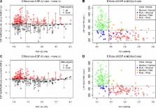
          When using three different immunoassays in cases with subjective cognitive decline and mild cognitive impairment, the CSF A β42/A β40 and A β42/A β38 ratios were significantly better predictors of abnormal amyloid PET than CSF A β42. Lower A β42, A β42/A β40, and A β42/A β38 ratios, but not A β40 and A β38, correlated with smaller hippocampal volumes measured by magnetic resonance imaging. However, lower A β38, A β40, and A β42, but not the ratios, correlated with non‐ AD‐specific subcortical changes, that is, larger lateral ventricles and white matter lesions. Further, the A β42/A β40 and A β42/A β38 ratios showed increased accuracy compared to A β42 when distinguishing AD from dementia with Lewy bodies or Parkinson's disease dementia and subcortical vascular dementia, where all A βs (including A β42) were decreased.

          Interpretation

          The CSF A β42/A β40 and A β42/A β38 ratios are significantly better than CSF A β42 to detect brain amyloid deposition in prodromal AD and to differentiate AD dementia from non‐ AD dementias. The ratios reflect AD‐type pathology better, whereas decline in CSF A β42 is also associated with non‐AD subcortical pathologies. These findings strongly suggest that the ratios rather than CSF A β42 should be used in the clinical work‐up of AD.

          Related collections

          Most cited references18

          • Record: found
          • Abstract: found
          • Article: not found

          APP processing and synaptic function.

          A large body of evidence has implicated Abeta peptides and other derivatives of the amyloid precursor protein (APP) as central to the pathogenesis of Alzheimer's disease (AD). However, the functional relationship of APP and its proteolytic derivatives to neuronal electrophysiology is not known. Here, we show that neuronal activity modulates the formation and secretion of Abeta peptides in hippocampal slice neurons that overexpress APP. In turn, Abeta selectively depresses excitatory synaptic transmission onto neurons that overexpress APP, as well as nearby neurons that do not. This depression depends on NMDA-R activity and can be reversed by blockade of neuronal activity. Synaptic depression from excessive Abeta could contribute to cognitive decline during early AD. In addition, we propose that activity-dependent modulation of endogenous Abeta production may normally participate in a negative feedback that could keep neuronal hyperactivity in check. Disruption of this feedback system could contribute to disease progression in AD.
            Bookmark
            • Record: found
            • Abstract: found
            • Article: not found

            Visualization of A beta 42(43) and A beta 40 in senile plaques with end-specific A beta monoclonals: evidence that an initially deposited species is A beta 42(43).

            To learn about the carboxy-terminal extent of amyloid beta-protein (A beta) composition of senile plaques (SPs) in the brain affected with Alzheimer's disease (AD), we employed two end-specific monoclonal antibodies as immunocytochemical probes: one is specific for A beta 40, the carboxyl terminus of A beta 1-40, while the other is specific for A beta 42(43). In the AD cortex, all SPs that were labeled with an authentic antibody were A beta 42(43) positive, while only one-third of which, on the average, were A beta 40 positive. There was a strong correlation between A beta 40 positivity and mature plaques. Two familial AD cortices with the mutation of beta-amyloid protein precursor 717 (beta APP717) (Val to Ile) showed a remarkable predominance of A beta 42(43)-positive, A beta 40-negative plaques. Diffuse plaques, representing the earliest stage of A beta deposition, were exclusively positive for A beta 42(43), but completely negative for A beta 40.
              Bookmark
              • Record: found
              • Abstract: found
              • Article: not found

              Comparing positron emission tomography imaging and cerebrospinal fluid measurements of β-amyloid.

              We examined agreement and disagreement between 2 biomarkers of β-amyloid (Aβ) deposition (amyloid positron emission tomography [PET] and cerebrospinal fluid [CSF] Aβ1-42 ) in normal aging and dementia in a large multicenter study. Concurrently acquired florbetapir PET and CSF Aβ were measured in cognitively normal, mild cognitive impairment (MCI), and Alzheimer's disease participants (n = 374) from the Alzheimer's Disease Neuroimaging Initiative. We also compared Aβ measurements in a separate group with serial CSF measurements over 3.1 ± 0.8 years that preceded a single florbetapir session. Additional biomarker and cognitive data allowed us to further examine profiles of discordant cases. Florbetapir and CSF Aβ were inversely correlated across all diagnostic groups, and dichotomous measurements were in agreement in 86% of subjects. Among subjects showing the most disagreement, the 2 discordant groups had different profiles: the florbetapir(+) /CSF Aβ(-) group was larger (n = 13) and was made up of only normal and early MCI subjects, whereas the florbetapir(-) /CSF Aβ(+) group was smaller (n = 7) and had poorer cognitive function and higher CSF tau, but no ApoE4 carriers. In the longitudinal sample, we observed both stable longitudinal CSF Aβ trajectories and those actively transitioning from normal to abnormal, but the final CSF Aβ measurements were in good agreement with florbetapir cortical retention. CSF and amyloid PET measurements of Aβ were consistent in the majority of subjects in the cross-sectional and longitudinal populations. Based on our analysis of discordant subjects, the available evidence did not show that CSF Aβ regularly becomes abnormal prior to fibrillar Aβ accumulation early in the course of disease. © 2013 American Neurological Association.
                Bookmark

                Author and article information

                Journal
                Ann Clin Transl Neurol
                Ann Clin Transl Neurol
                10.1002/(ISSN)2328-9503
                ACN3
                Annals of Clinical and Translational Neurology
                John Wiley and Sons Inc. (Hoboken )
                2328-9503
                01 January 2016
                March 2016
                : 3
                : 3 ( doiID: 10.1111/acn3.2016.3.issue-3 )
                : 154-165
                Affiliations
                [ 1 ] Clinical Memory Research Unit Department of Clinical Sciences, MalmöLund University LundSweden
                [ 2 ] Clinical Neurochemistry Laboratory Institute of Neuroscience and PhysiologySahlgrenska Academy at the University of Gothenburg MölndalSweden
                [ 3 ] Department of Molecular NeuroscienceUCL Institute of Neurology Queen Square LondonUnited Kingdom
                [ 4 ] Memory ClinicSkåne University Hospital MalmöSweden
                [ 5 ] Department of NeurologySkåne University Hospital MalmöSweden
                [ 6 ]ADx NeuroSciences GentBelgium
                [ 7 ] Department of Clinical Sciences Diagnostic RadiologyLund University LundSweden
                [ 8 ] Imaging and FunctionSkåne University Health Care LundSweden
                Author notes
                [*] [* ] Correspondence

                Shorena Janelidze, Clinical Memory Research Unit, Department of Clinical Sciences Malmö, SE‐221 00 Lund, Sweden. Tel: +46 46‐2229667; Fax: +46 40‐335657; E‐mail: Shorena.Janelidze@ 123456med.lu.se

                Oskar Hansson, Memory Clinic, Skåne University Hospital, SE‐20502 Malmö, Sweden. Tel: +46 40‐335036; Fax: +46 40‐335657; E‐mail: Oskar.Hansson@ 123456med.lu.se

                Article
                ACN3274
                10.1002/acn3.274
                4774260
                27042676
                26134ffe-e6f0-4a1b-bd9f-edc3409c476b
                © 2016 The Authors. Annals of Clinical and Translational Neurology published by Wiley Periodicals, Inc on behalf of American Neurological Association.

                This is an open access article under the terms of the Creative Commons Attribution‐NonCommercial‐NoDerivs License, which permits use and distribution in any medium, provided the original work is properly cited, the use is non‐commercial and no modifications or adaptations are made.

                History
                : 20 October 2015
                : 13 November 2015
                : 16 November 2015
                Page count
                Pages: 12
                Funding
                Funded by: European Research Council
                Funded by: Swedish Research Council
                Funded by: Strategic Research Area MultiPark (Multidisciplinary Research in Parkinson's disease) at Lund University
                Funded by: Crafoord Foundation
                Funded by: Swedish Brain Foundation
                Funded by: Swedish Alzheimer Foundation
                Funded by: Torsten Söderberg Foundation
                Funded by: Knut and Alice Wallenberg Foundation
                Funded by: Swedish Federal Government
                Categories
                Research Article
                Research Articles
                Custom metadata
                2.0
                acn3274
                March 2016
                Converter:WILEY_ML3GV2_TO_NLMPMC version:4.7.6 mode:remove_FC converted:02.03.2016

                Comments

                Comment on this article