5
views
0
recommends
+1 Recommend
0 collections
    0
    shares
      • Record: found
      • Abstract: found
      • Article: found
      Is Open Access

      The Structure and Function of Acylglycerophosphate Acyltransferase 4/ Lysophosphatidic Acid Acyltransferase Delta (AGPAT4/LPAATδ)

      review-article

      Read this article at

      Bookmark
          There is no author summary for this article yet. Authors can add summaries to their articles on ScienceOpen to make them more accessible to a non-specialist audience.

          Abstract

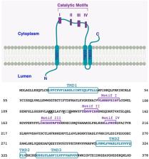
          Lipid-modifying enzymes serve crucial roles in cellular processes such as signal transduction (producing lipid-derived second messengers), intracellular membrane transport (facilitating membrane remodeling needed for membrane fusion/fission), and protein clustering (organizing lipid domains as anchoring platforms). The lipid products crucial in these processes can derive from different metabolic pathways, thus it is essential to know the localization, substrate specificity, deriving products (and their function) of all lipid-modifying enzymes. Here we discuss an emerging family of these enzymes, the lysophosphatidic acid acyltransferases (LPAATs), also known as acylglycerophosphate acyltransferases (AGPATs), that produce phosphatidic acid (PA) having as substrates lysophosphatidic acid (LPA) and acyl-CoA. Eleven LPAAT/AGPAT enzymes have been identified in mice and humans based on sequence homologies, and their localization, specific substrates and functions explored. We focus on one member of the family, LPAATδ, a protein expressed mainly in brain and in muscle (though to a lesser extent in other tissues); while at the cellular level it is localized at the trans-Golgi network membranes and at the mitochondrial outer membranes. LPAATδ is a physiologically essential enzyme since mice knocked-out for Lpaatδ show severe dysfunctions including cognitive impairment, impaired force contractility and altered white adipose tissue. The LPAATδ physiological roles are related to the formation of its product PA. PA is a multifunctional lipid involved in cell signaling as well as in membrane remodeling. In particular, the LPAATδ-catalyzed conversion of LPA (inverted-cone-shaped lipid) to PA (cone-shaped lipid) is considered a mechanism of deformation of the bilayer that favors membrane fission. Indeed, LPAATδ is an essential component of the fission-inducing machinery driven by the protein BARS. In this process, a protein-tripartite complex (BARS/14-3-3γ/phosphoinositide kinase PI4KIIIβ) is recruited at the trans-Golgi network, at the sites where membrane fission is to occur; there, LPAATδ directly interacts with BARS and is activated by BARS. The resulting formation of PA is essential for membrane fission occurring at those spots. Also in mitochondria PA formation has been related to fusion/fission events. Since PA is formed by various enzymatic pathways in different cell compartments, the BARS-LPAATδ interaction indicates the relevance of lipid-modifying enzymes acting exactly where their products are needed (i.e., PA at the Golgi membranes).

          Related collections

          Most cited references103

          • Record: found
          • Abstract: found
          • Article: not found

          How lipids affect the activities of integral membrane proteins.

          The activities of integral membrane proteins are often affected by the structures of the lipid molecules that surround them in the membrane. One important parameter is the hydrophobic thickness of the lipid bilayer, defined by the lengths of the lipid fatty acyl chains. Membrane proteins are not rigid entities, and deform to ensure good hydrophobic matching to the surrounding lipid bilayer. The structure of the lipid headgroup region is likely to be important in defining the structures of those parts of a membrane protein that are located in the lipid headgroup region. A number of examples are given where the conformation of the headgroup-embedded region of a membrane protein changes during the reaction cycle of the protein; activities of such proteins might be expected to be particularly sensitive to lipid headgroup structure. Differences in hydrogen bonding potential and hydration between the headgroups of phosphatidycholines and phosphatidylethanolamines could be important factors in determining the effects of these lipids on protein activities, as well as any effects related to the tendency of the phosphatidylethanolamines to form a curved, hexagonal H(II) phase. Effects of lipid structure on protein aggregation and helix-helix interactions are also discussed, as well as the effects of charged lipids on ion concentrations close to the surface of the bilayer. Interpretations of lipid effects in terms of changes in protein volume, lipid free volume, and curvature frustration are also described. Finally, the role of non-annular, or 'co-factor' lipids, tightly bound to membrane proteins, is described.
            Bookmark
            • Record: found
            • Abstract: not found
            • Article: not found

            The function of cytidine coenzymes in the biosynthesis of phospholipides.

              Bookmark
              • Record: found
              • Abstract: found
              • Article: not found

              Protein-lipid interplay in fusion and fission of biological membranes.

              Disparate biological processes involve fusion of two membranes into one and fission of one membrane into two. To formulate the possible job description for the proteins that mediate remodeling of biological membranes, we analyze the energy price of disruption and bending of membrane lipid bilayers at the different stages of bilayer fusion. The phenomenology and the pathways of the well-characterized reactions of biological remodeling, such as fusion mediated by influenza hemagglutinin, are compared with those studied for protein-free bilayers. We briefly consider some proteins involved in fusion and fission, and the dependence of remodeling on the lipid composition of the membranes. The specific hypothetical mechanisms by which the proteins can lower the energy price of the bilayer rearrangement are discussed in light of the experimental data and the requirements imposed by the elastic properties of the bilayer.
                Bookmark

                Author and article information

                Contributors
                Journal
                Front Cell Dev Biol
                Front Cell Dev Biol
                Front. Cell Dev. Biol.
                Frontiers in Cell and Developmental Biology
                Frontiers Media S.A.
                2296-634X
                02 August 2019
                2019
                : 7
                : 147
                Affiliations
                Institute of Biochemistry and Cell Biology and Institute of Protein Biochemistry, National Research Council , Naples, Italy
                Author notes

                Edited by: Vladimir Lupashin, University of Arkansas for Medical Sciences, United States

                Reviewed by: Mitsuo Tagaya, Tokyo University of Pharmacy and Life Sciences, Japan; Robin Duncan, University of Waterloo, Canada

                *Correspondence: Mikhail A. Zhukovsky, m.zhukovsky@ 123456ibp.cnr.it

                This article was submitted to Membrane Traffic, a section of the journal Frontiers in Cell and Developmental Biology

                Article
                10.3389/fcell.2019.00147
                6688108
                31428612
                24d40c89-9c9a-4471-8740-9a209be3400b
                Copyright © 2019 Zhukovsky, Filograna, Luini, Corda and Valente.

                This is an open-access article distributed under the terms of the Creative Commons Attribution License (CC BY). The use, distribution or reproduction in other forums is permitted, provided the original author(s) and the copyright owner(s) are credited and that the original publication in this journal is cited, in accordance with accepted academic practice. No use, distribution or reproduction is permitted which does not comply with these terms.

                History
                : 07 May 2019
                : 16 July 2019
                Page count
                Figures: 4, Tables: 0, Equations: 0, References: 114, Pages: 15, Words: 0
                Funding
                Funded by: Associazione Italiana per la Ricerca sul Cancro 10.13039/501100005010
                Award ID: IG17524
                Award ID: IG10341
                Award ID: IG18776
                Award ID: IG20786
                Award ID: IG15767
                Funded by: Ministero dell’Istruzione, dell’Università e della Ricerca 10.13039/501100003407
                Award ID: CNT01_00177_962865
                Categories
                Physiology
                Review

                acyltransferase,agpat,lpaat,phosphatidic acid,lysophosphatidic acid,golgi complex,membrane fission,bars

                Comments

                Comment on this article