31
views
0
recommends
+1 Recommend
0 collections
    0
    shares
      • Record: found
      • Abstract: found
      • Article: found
      Is Open Access

      The Dual Nature of Type I and Type II Interferons

      review-article

      Read this article at

      Bookmark
          There is no author summary for this article yet. Authors can add summaries to their articles on ScienceOpen to make them more accessible to a non-specialist audience.

          Abstract

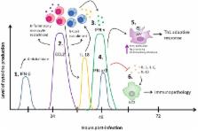
          Type I and type II interferons (IFN) are central to both combating virus infection and modulating the antiviral immune response. Indeed, an absence of either the receptor for type I IFNs or IFN-y have resulted in increased susceptibility to virus infection, including increased virus replication and reduced survival. However, an emerging area of research has shown that there is a dual nature to these cytokines. Recent evidence has demonstrated that both type I and type II IFNs have immunoregulatory functions during infection and type II immune responses. In this review, we address the dual nature of type I and type II interferons and present evidence that both antiviral and immunomodulatory functions are critical during virus infection to not only limit virus replication and initiate an appropriate antiviral immune response, but to also negatively regulate this response to minimize tissue damage. Both the activating and negatively regulatory properties of type I and II IFNs work in concert with each other to create a balanced immune response that combats the infection while minimizing collateral damage.

          Related collections

          Most cited references70

          • Record: found
          • Abstract: found
          • Article: not found

          Nitric oxide and macrophage function.

          At the interface between the innate and adaptive immune systems lies the high-output isoform of nitric oxide synthase (NOS2 or iNOS). This remarkable molecular machine requires at least 17 binding reactions to assemble a functional dimer. Sustained catalysis results from the ability of NOS2 to attach calmodulin without dependence on elevated Ca2+. Expression of NOS2 in macrophages is controlled by cytokines and microbial products, primarily by transcriptional induction. NOS2 has been documented in macrophages from human, horse, cow, goat, sheep, rat, mouse, and chicken. Human NOS2 is most readily observed in monocytes or macrophages from patients with infectious or inflammatory diseases. Sustained production of NO endows macrophages with cytostatic or cytotoxic activity against viruses, bacteria, fungi, protozoa, helminths, and tumor cells. The antimicrobial and cytotoxic actions of NO are enhanced by other macrophage products such as acid, glutathione, cysteine, hydrogen peroxide, or superoxide. Although the high-output NO pathway probably evolved to protect the host from infection, suppressive effects on lymphocyte proliferation and damage to other normal host cells confer upon NOS2 the same protective/destructive duality inherent in every other major component of the immune response.
            Bookmark
            • Record: found
            • Abstract: found
            • Article: not found

            The IRF family transcription factors in immunity and oncogenesis.

            The interferon regulatory factor (IRF) family, consisting of nine members in mammals, was identified in the late 1980s in the context of research into the type I interferon system. Subsequent studies over the past two decades have revealed the versatile and critical functions performed by this transcription factor family. Indeed, many IRF members play central roles in the cellular differentiation of hematopoietic cells and in the regulation of gene expression in response to pathogen-derived danger signals. In particular, the advances made in understanding the immunobiology of Toll-like and other pattern-recognition receptors have recently generated new momentum for the study of IRFs. Moreover, the role of several IRF family members in the regulation of the cell cycle and apoptosis has important implications for understanding susceptibility to and progression of several cancers.
              Bookmark
              • Record: found
              • Abstract: found
              • Article: found
              Is Open Access

              Anifrolumab, an Anti–Interferon‐α Receptor Monoclonal Antibody, in Moderate‐to‐Severe Systemic Lupus Erythematosus

              Objective To assess the efficacy and safety of anifrolumab, a type I interferon (IFN) receptor antagonist, in a phase IIb, randomized, double‐blind, placebo‐controlled study of adults with moderate‐to‐severe systemic lupus erythematosus (SLE). Methods Patients (n = 305) were randomized to receive intravenous anifrolumab (300 mg or 1,000 mg) or placebo, in addition to standard therapy, every 4 weeks for 48 weeks. Randomization was stratified by SLE Disease Activity Index 2000 score (<10 or ≥10), oral corticosteroid dosage (<10 or ≥10 mg/day), and type I IFN gene signature test status (high or low) based on a 4‐gene expression assay. The primary end point was the percentage of patients achieving an SLE Responder Index (SRI[4]) response at week 24 with sustained reduction of oral corticosteroids (<10 mg/day and less than or equal to the dose at week 1 from week 12 through 24). Other end points (including SRI[4], British Isles Lupus Assessment Group [BILAG]–based Composite Lupus Assessment [BICLA], modified SRI[6], and major clinical response) were assessed at week 52. The primary end point was analyzed in the modified intent‐to‐treat (ITT) population and type I IFN–high subpopulation. The study result was considered positive if the primary end point was met in either of the 2 study populations. The Type I error rate was controlled at 0.10 (2‐sided), within each of the 2 study populations for the primary end point analysis. Results The primary end point was met by more patients treated with anifrolumab (34.3% of 99 for 300 mg and 28.8% of 104 for 1,000 mg) than placebo (17.6% of 102) (P = 0.014 for 300 mg and P = 0.063 for 1,000 mg, versus placebo), with greater effect size in patients with a high IFN signature at baseline (13.2% in placebo‐treated patients versus 36.0% [P = 0.004] and 28.2% [P = 0.029]) in patients treated with anifrolumab 300 mg and 1,000 mg, respectively. At week 52, patients treated with anifrolumab achieved greater responses in SRI(4) (40.2% versus 62.6% [P < 0.001] and 53.8% [P = 0.043] with placebo, anifrolumab 300 mg, and anifrolumab 1,000 mg, respectively), BICLA (25.7% versus 53.5% [P < 0.001] and 41.2% [P = 0.018], respectively), modified SRI(6) (28.4% versus 49.5% [P = 0.002] and 44.7% [P = 0.015], respectively), major clinical response (BILAG 2004 C or better in all organ domains from week 24 through week 52) (6.9% versus 19.2% [P = 0.012] and 17.3% [P = 0.025], respectively), and several other global and organ‐specific end points. Herpes zoster was more frequent in the anifrolumab‐treated patients (2.0% with placebo treatment versus 5.1% and 9.5% with anifrolumab 300 mg and 1,000 mg, respectively), as were cases reported as influenza (2.0% versus 6.1% and 7.6%, respectively), in the anifrolumab treatment groups. Incidence of serious adverse events was similar between groups (18.8% versus 16.2% and 17.1%, respectively). Conclusion Anifrolumab substantially reduced disease activity compared with placebo across multiple clinical end points in the patients with moderate‐to‐severe SLE.
                Bookmark

                Author and article information

                Contributors
                Journal
                Front Immunol
                Front Immunol
                Front. Immunol.
                Frontiers in Immunology
                Frontiers Media S.A.
                1664-3224
                11 September 2018
                2018
                : 9
                : 2061
                Affiliations
                Department of Pathology and Molecular Medicine, McMaster Immunology Research Centre, McMaster University , Hamilton, ON, Canada
                Author notes

                Edited by: Maziar Divangahi, McGill University, Canada

                Reviewed by: Jianzhong Zhu, Yangzhou University, China; Alain Lamarre, Institut National de la Recherche Scientifique (INRS), Canada

                *Correspondence: Ali A. Ashkar ashkara@ 123456mcmaster.ca

                This article was submitted to Viral Immunology, a section of the journal Frontiers in Immunology

                Article
                10.3389/fimmu.2018.02061
                6141705
                30254639
                249eeedd-280e-4a37-aa3c-805d7c8d8c2c
                Copyright © 2018 Lee and Ashkar.

                This is an open-access article distributed under the terms of the Creative Commons Attribution License (CC BY). The use, distribution or reproduction in other forums is permitted, provided the original author(s) and the copyright owner(s) are credited and that the original publication in this journal is cited, in accordance with accepted academic practice. No use, distribution or reproduction is permitted which does not comply with these terms.

                History
                : 01 June 2018
                : 21 August 2018
                Page count
                Figures: 1, Tables: 2, Equations: 0, References: 98, Pages: 10, Words: 8367
                Categories
                Immunology
                Review

                Immunology
                type i interferon,interferon-γ,innate immunity,virus infection,immunoregulation
                Immunology
                type i interferon, interferon-γ, innate immunity, virus infection, immunoregulation

                Comments

                Comment on this article