13
views
0
recommends
+1 Recommend
0 collections
    0
    shares
      • Record: found
      • Abstract: found
      • Article: found
      Is Open Access

      Integration of Brain and Skull in Prenatal Mouse Models of Apert and Crouzon Syndromes

      research-article

      Read this article at

      Bookmark
          There is no author summary for this article yet. Authors can add summaries to their articles on ScienceOpen to make them more accessible to a non-specialist audience.

          Abstract

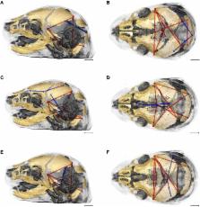
          The brain and skull represent a complex arrangement of integrated anatomical structures composed of various cell and tissue types that maintain structural and functional association throughout development. Morphological integration, a concept developed in vertebrate morphology and evolutionary biology, describes the coordinated variation of functionally and developmentally related traits of organisms. Syndromic craniosynostosis is characterized by distinctive changes in skull morphology and perceptible, though less well studied, changes in brain structure and morphology. Using mouse models for craniosynostosis conditions, our group has precisely defined how unique craniosynostosis causing mutations in fibroblast growth factor receptors affect brain and skull morphology and dysgenesis involving coordinated tissue-specific effects of these mutations. Here we examine integration of brain and skull in two mouse models for craniosynostosis: one carrying the FGFR2c C342Y mutation associated with Pfeiffer and Crouzon syndromes and a mouse model carrying the FGFR2 S252W mutation, one of two mutations responsible for two-thirds of Apert syndrome cases. Using linear distances estimated from three-dimensional coordinates of landmarks acquired from dual modality imaging of skull (high resolution micro-computed tomography and magnetic resonance microscopy) of mice at embryonic day 17.5, we confirm variation in brain and skull morphology in Fgfr2c C342Y/+ mice, Fgfr2 +/ S252W mice, and their unaffected littermates. Mutation-specific variation in neural and cranial tissue notwithstanding, patterns of integration of brain and skull differed only subtly between mice carrying either the FGFR2c C342Y or the FGFR2 S252W mutation and their unaffected littermates. However, statistically significant and substantial differences in morphological integration of brain and skull were revealed between the two mutant mouse models, each maintained on a different strain. Relative to the effects of disease-associated mutations, our results reveal a stronger influence of the background genome on patterns of brain-skull integration and suggest robust genetic, developmental, and evolutionary relationships between neural and skeletal tissues of the head.

          Related collections

          Most cited references84

          • Record: found
          • Abstract: not found
          • Article: not found

          Morphological Integration and Developmental Modularity

            Bookmark
            • Record: found
            • Abstract: not found
            • Article: not found

            FGF signaling pathways in endochondral and intramembranous bone development and human genetic disease.

              Bookmark
              • Record: found
              • Abstract: found
              • Article: not found

              Deciphering the Palimpsest: Studying the Relationship Between Morphological Integration and Phenotypic Covariation.

              Organisms represent a complex arrangement of anatomical structures and individuated parts that must maintain functional associations through development. This integration of variation between functionally related body parts and the modular organization of development are fundamental determinants of their evolvability. This is because integration results in the expression of coordinated variation that can create preferred directions for evolutionary change, while modularity enables variation in a group of traits or regions to accumulate without deleterious effects on other aspects of the organism. Using our own work on both model systems (e.g., lab mice, avians) and natural populations of rodents and primates, we explore in this paper the relationship between patterns of phenotypic covariation and the developmental determinants of integration that those patterns are assumed to reflect. We show that integration cannot be reliably studied through phenotypic covariance patterns alone and argue that the relationship between phenotypic covariation and integration is obscured in two ways. One is the superimposition of multiple determinants of covariance in complex systems and the other is the dependence of covariation structure on variances in covariance-generating processes. As a consequence, we argue that the direct study of the developmental determinants of integration in model systems is necessary to fully interpret patterns of covariation in natural populations, to link covariation patterns to the processes that generate them, and to understand their significance for evolutionary explanation.
                Bookmark

                Author and article information

                Contributors
                Journal
                Front Hum Neurosci
                Front Hum Neurosci
                Front. Hum. Neurosci.
                Frontiers in Human Neuroscience
                Frontiers Media S.A.
                1662-5161
                25 July 2017
                2017
                : 11
                : 369
                Affiliations
                [1] 1Department of Anthropology, Pennsylvania State University University Park, PA, United States
                [2] 2Center for Quantitative Imaging, Penn State Institutes for Energy and the Environment, Pennsylvania State University University Park, PA, United States
                [3] 3High Field MRI Facility, Huck Institutes of the Life Sciences, Pennsylvania State University University Park, PA, United States
                [4] 4Department of Bioengineering, Pennsylvania State University University Park, PA, United States
                [5] 5Department of Genetics and Genomic Sciences, Icahn School of Medicine at Mount Sinai New York, NY, United States
                Author notes

                Edited by: Wanda Lattanzi, Università Cattolica Del Sacro Cuore, Italy

                Reviewed by: Natalina Quarto, University of Naples Federico II, Italy; Mark Harold Moore, Australian Craniofacial Unit, Australia

                *Correspondence: Joan T. Richtsmeier jta10@ 123456psu.edu

                †These authors have contributed equally to this work.

                Article
                10.3389/fnhum.2017.00369
                5525342
                28790902
                24884b16-8a88-4c36-bd7b-66699c785671
                Copyright © 2017 Motch Perrine, Stecko, Neuberger, Jabs, Ryan and Richtsmeier.

                This is an open-access article distributed under the terms of the Creative Commons Attribution License (CC BY). The use, distribution or reproduction in other forums is permitted, provided the original author(s) or licensor are credited and that the original publication in this journal is cited, in accordance with accepted academic practice. No use, distribution or reproduction is permitted which does not comply with these terms.

                History
                : 16 May 2017
                : 29 June 2017
                Page count
                Figures: 7, Tables: 2, Equations: 0, References: 90, Pages: 15, Words: 11502
                Funding
                Funded by: National Institute of Child Health and Human Development 10.13039/100000071
                Award ID: P01HD078233
                Funded by: National Institute of Dental and Craniofacial Research 10.13039/100000072
                Award ID: R01-DE018500
                Award ID: R01-DE022988
                Award ID: R01-DE018500-S1
                Categories
                Neuroscience
                Original Research

                Neurosciences
                morphological integration,brain,skull,crouzon syndrome,apert syndrome,craniosynostosis,development,craniofacial

                Comments

                Comment on this article