31
views
0
recommends
+1 Recommend
0 collections
    0
    shares
      • Record: found
      • Abstract: found
      • Article: found
      Is Open Access

      Individual Trabeculae Segmentation (ITS)–Based Morphological Analysis of High-Resolution Peripheral Quantitative Computed Tomography Images Detects Abnormal Trabecular Plate and Rod Microarchitecture in Premenopausal Women With Idiopathic Osteoporosis

      research-article

      Read this article at

      Bookmark
          There is no author summary for this article yet. Authors can add summaries to their articles on ScienceOpen to make them more accessible to a non-specialist audience.

          Abstract

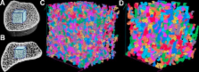
          Idiopathic osteoporosis (IOP) in premenopausal women is a poorly understood entity in which otherwise healthy women have low-trauma fracture or very low bone mineral density (BMD). In this study, we applied individual trabeculae segmentation (ITS)–based morphological analysis to high-resolution peripheral quantitative computed tomography (HR-pQCT) images of the distal radius and distal tibia to gain greater insight into skeletal microarchitecture in premenopausal women with IOP. HR-pQCT scans were performed for 26 normal control individuals and 31 women with IOP. A cubic subvolume was extracted from the trabecular bone compartment and subjected to ITS-based analysis. Three Young's moduli and three shear moduli were calculated by micro–finite element (µFE) analysis. ITS-based morphological analysis of HR-pQCT images detected significantly decreased trabecular plate and rod bone volume fraction and number, decreased axial bone volume fraction in the longitudinal axis, increased rod length, and decreased rod-to-rod, plate-to-rod, and plate-to-plate junction densities at the distal radius and distal tibia in women with IOP. However, trabecular plate and rod thickness did not differ. A more rod-like trabecular microstructure was found in the distal radius, but not in the distal tibia. Most ITS measurements contributed significantly to the elastic moduli of trabecular bone independent of bone volume fraction (BV/TV). At a fixed BV/TV, plate-like trabeculae contributed positively to the mechanical properties of trabecular bone. The results suggest that ITS-based morphological analysis of HR-pQCT images is a sensitive and promising clinical tool for the investigation of trabecular bone microstructure in human studies of osteoporosis. © 2010 American Society for Bone and Mineral Research.

          Related collections

          Most cited references49

          • Record: found
          • Abstract: found
          • Article: not found

          In vivo assessment of trabecular bone microarchitecture by high-resolution peripheral quantitative computed tomography.

          Assessment of trabecular microarchitecture may enhance the prediction of fracture risk and improve monitoring of treatment response. A new high-resolution peripheral quantitative computed tomography (HR-pQCT) system permits in vivo assessment of trabecular architecture and volumetric bone mineral density (BMD) at the distal radius and tibia with a voxel size of 82 microm3. We determined the short-term reproducibility of this device by measuring 15 healthy volunteers three times each. We compared HR-pQCT measurements in 108 healthy premenopausal, 113 postmenopausal osteopenic, and 35 postmenopausal osteoporotic women. Furthermore, we compared values in postmenopausal osteopenic women with (n = 35) and without previous fracture history (n = 78). We conducted a cross-sectional study in a private clinical research center. We took HR-pQCT measurements of the radius and tibia. Femoral neck and spine BMD were measured in postmenopausal women by dual-energy x-ray absorptiometry. Precision of HR-pQCT measurements was 0.7-1.5% for total, trabecular, and cortical densities and 2.5-4.4% for trabecular architecture. Postmenopausal women had lower density, trabecular number, and cortical thickness than premenopausal women (P < 0.001) at both radius and tibia. Osteoporotic women had lower density, cortical thickness, and increased trabecular separation than osteopenic women (P < 0.01) at both sites. Furthermore, although spine and hip BMD were similar, fractured osteopenic women had lower trabecular density and more heterogeneous trabecular distribution (P < 0.02) at the radius compared with unfractured osteopenic women. HR-pQCT appears promising to assess bone density and microarchitecture at peripheral sites in terms of reproducibility and ability to detect age- and disease-related changes.
            Bookmark
            • Record: found
            • Abstract: found
            • Article: not found

            Quantification of Bone Microarchitecture with the Structure Model Index.

            The deterioration of cancellous bone structure due to aging and disease is characterized by a conversion from plate elements to rod elements. Consequently the terms "rod-like" and "plate-like" are frequently used for a subjective classification of cancellous bone. In this work a new morphometric parameter called Structure Model Index (SMI) is introduced, which makes it possible to quantify the characteristic form of a three-dimensionally described structure in terms of the amount of plates and rod composing the structure. The SMI is calculated by means of three-dimensional image analysis based on a differential analysis of the triangulated bone surface. For an ideal plate and rod structure the SMI value is 0 and 3, respectively, independent of the physical dimensions. For a structure with both plates and rods of equal thickness the value lies between 0 and 3, depending on the volume ratio of rods and plates. The SMI parameter is evaluated by examining bone biopsies from different skeletal sites. The bone samples were measured three-dimensionally with a micro-CT system. Samples with the same volume density but varying trabecular architecture can uniquely be characterized with the SMI. Furthermore the SMI values were found to correspond well with the perceived structure type.
              Bookmark
              • Record: found
              • Abstract: found
              • Article: not found

              Relationships between surface, volume, and thickness of iliac trabecular bone in aging and in osteoporosis. Implications for the microanatomic and cellular mechanisms of bone loss.

              We devised a new method for examining the structural changes that occur in trabecular bone in aging and in osteoporosis. With simultaneous measurement of total perimeter and bone area in thin sections, indirect indices of mean trabecular plate thickness (MTPT) and mean trabecular plate density (MTPD) can be derived, such that trabecular bone volume = MTPD X MTPT. MTPD is an index of the probability that a scanning or test line will intersect a structural element of bone, and is the reciprocal of the mean distance between the midpoints of structural elements, multiplied by pi/2. We applied this method to iliac bone samples from 78 normal subjects, 100 patients with vertebral fracture, and 50 patients with hip fracture. The reduction in trabecular bone volume observed in normal subjects with increasing age was mainly due to a reduction in plate density, with no significant decrease in plate thickness. The further reduction in trabecular bone volume observed in patients with osteoporotic vertebral fracture was mainly due to a further reduction in plate density. There was a relatively smaller reduction in plate thickness that was statistically significant in males but not in females. Only in patients with hip fracture did trabecular thinning contribute substantially to the additional loss of trabecular bone in osteoporosis relative to age. These data indicate that age-related bone loss occurs principally by a process that removes entire structural elements of bone; those that remain are more widely separated and some may undergo compensatory thickening, but most slowly become reduced in thickness. We propose that the process of removal is initiated by increased depth of osteoclastic resorption cavities which leads to focal perforation of trabecular plates; this is followed by progressive enlargement of the perforations with conversion of plates to rods. The resulting structural changes are more severe in osteoporotic patients than in normal subjects, but have been completed in most patients before they develop symptoms.
                Bookmark

                Author and article information

                Journal
                J Bone Miner Res
                jbmr
                Journal of Bone and Mineral Research
                Wiley Subscription Services, Inc., A Wiley Company
                0884-0431
                1523-4681
                July 2010
                25 January 2010
                : 25
                : 7
                : 1496-1505
                Affiliations
                [1 ]simpleBone Bioengineering Laboratory, Department of Biomedical Engineering, Columbia University New York, NY, USA
                [2 ]simpleDivision of Endocrinology, Department of Medicine, Columbia University New York, NY, USA
                [3 ]simpleDepartment of Medicine, Osteoporosis Research Center, Creighton University Omaha, NB, USA
                Author notes
                Address correspondence to: X Edward Guo, PhD, Department of Biomedical Engineering, Columbia University, 351 Engineering Terrace, Mail Code 8904, 1210 Amsterdam Avenue, New York, NY 10027, USA. E-mail: ed.guo@ 123456columbia.edu
                Article
                10.1002/jbmr.50
                3131618
                20200967
                246244cc-2a19-4d10-b788-88df8f54cb3e
                Copyright © 2010 American Society for Bone and Mineral Research

                Re-use of this article is permitted in accordance with the Creative Commons Deed, Attribution 2.5, which does not permit commercial exploitation.

                History
                : 30 June 2009
                : 26 August 2009
                : 19 January 2010
                Categories
                Original Article

                Human biology
                bone microstructure,trabecular plate/rod,high-resolution peripheral quantitative computed tomography,individual trabeculae segmentation

                Comments

                Comment on this article