9
views
0
recommends
+1 Recommend
0 collections
    0
    shares
      • Record: found
      • Abstract: found
      • Article: found
      Is Open Access

      Growth Differentiation Factor 15 Regulates Oxidative Stress-Dependent Ferroptosis Post Spinal Cord Injury by Stabilizing the p62-Keap1-Nrf2 Signaling Pathway

      research-article

      Read this article at

      Bookmark
          There is no author summary for this article yet. Authors can add summaries to their articles on ScienceOpen to make them more accessible to a non-specialist audience.

          Abstract

          Background

          Spinal cord injury (SCI) is a severe traumatic disorder of the central nervous system (CNS) that causes irreversible damage to the nervous tissue. The consequent hemorrhage contributed by trauma induces neuronal ferroptosis post SCI, which is an important death mode to mediate neuronal loss. Growth differentiation factor 15 (GDF15) is a cytokine that regulates cell proliferation, differentiation, and death. However, the specific role of GDF15 in neuronal ferroptosis post SCI remains unknown.

          Materials and Methods

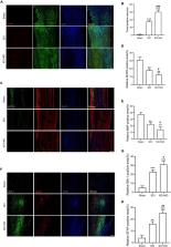
          Neuronal ferroptosis in vitro was measured by detection of lipid peroxidation, glutathione, iron content, and reactive oxidative stress. In vivo, western blotting and immunofluorescence (IF) staining was utilized to measure ferroptosis post SCI. IF staining, TUNEL staining, hematoxylin-eosin staining, and Nissl staining were used to measure neurological damage. Finally, locomotor function recovery was analyzed using the Basso Mouse Scale and Louisville Swim Scale.

          Results

          GDF15 was significantly increased in neuronal ferroptosis and silencing GDF15 aggravated ferroptosis both in vitro and in vivo. Besides, GDF15-mediated inhibition of neuronal ferroptosis is through p62-dependent Keap1-Nrf2 pathway. In SCI mice, knockdown of GDF15 significantly exacerbated neuronal death, interfered with axon regeneration and remyelination, aggravated ferroptosis-mediated neuroinflammation, and restrained locomotor recovery.

          Conclusion

          GDF15 effectively alleviated neuronal ferroptosis post SCI via the p62-Keap1-Nrf2 signaling pathway and promoted locomotor recovery of SCI mice, which is suggested as a potential target on SCI pathogenesis and treatment.

          Related collections

          Most cited references51

          • Record: found
          • Abstract: found
          • Article: not found

          Ferroptosis: an iron-dependent form of nonapoptotic cell death.

          Nonapoptotic forms of cell death may facilitate the selective elimination of some tumor cells or be activated in specific pathological states. The oncogenic RAS-selective lethal small molecule erastin triggers a unique iron-dependent form of nonapoptotic cell death that we term ferroptosis. Ferroptosis is dependent upon intracellular iron, but not other metals, and is morphologically, biochemically, and genetically distinct from apoptosis, necrosis, and autophagy. We identify the small molecule ferrostatin-1 as a potent inhibitor of ferroptosis in cancer cells and glutamate-induced cell death in organotypic rat brain slices, suggesting similarities between these two processes. Indeed, erastin, like glutamate, inhibits cystine uptake by the cystine/glutamate antiporter (system x(c)(-)), creating a void in the antioxidant defenses of the cell and ultimately leading to iron-dependent, oxidative death. Thus, activation of ferroptosis results in the nonapoptotic destruction of certain cancer cells, whereas inhibition of this process may protect organisms from neurodegeneration. Copyright © 2012 Elsevier Inc. All rights reserved.
            Bookmark
            • Record: found
            • Abstract: found
            • Article: not found

            Ferroptosis: Death by Lipid Peroxidation.

            Ferroptosis is a regulated form of cell death driven by loss of activity of the lipid repair enzyme glutathione peroxidase 4 (GPX4) and subsequent accumulation of lipid-based reactive oxygen species (ROS), particularly lipid hydroperoxides. This form of iron-dependent cell death is genetically, biochemically, and morphologically distinct from other cell death modalities, including apoptosis, unregulated necrosis, and necroptosis. Ferroptosis is regulated by specific pathways and is involved in diverse biological contexts. Here we summarize the discovery of ferroptosis, the mechanism of ferroptosis regulation, and its increasingly appreciated relevance to both normal and pathological physiology.
              Bookmark
              • Record: found
              • Abstract: found
              • Article: not found

              Activation of the p62-Keap1-NRF2 pathway protects against ferroptosis in hepatocellular carcinoma cells.

              Ferroptosis is a recently recognized form of regulated cell death caused by an iron-dependent accumulation of lipid reactive oxygen species. However, the molecular mechanisms regulating ferroptosis remain obscure. Here, we report that nuclear factor erythroid 2-related factor 2 (NRF2) plays a central role in protecting hepatocellular carcinoma (HCC) cells against ferroptosis. Upon exposure to ferroptosis-inducing compounds (e.g., erastin, sorafenib, and buthionine sulfoximine), p62 expression prevented NRF2 degradation and enhanced subsequent NRF2 nuclear accumulation through inactivation of Kelch-like ECH-associated protein 1. Additionally, nuclear NRF2 interacted with transcriptional coactivator small v-maf avian musculoaponeurotic fibrosarcoma oncogene homolog proteins such as MafG and then activated transcription of quinone oxidoreductase-1, heme oxygenase-1, and ferritin heavy chain-1. Knockdown of p62, quinone oxidoreductase-1, heme oxygenase-1, and ferritin heavy chain-1 by RNA interference in HCC cells promoted ferroptosis in response to erastin and sorafenib. Furthermore, genetic or pharmacologic inhibition of NRF2 expression/activity in HCC cells increased the anticancer activity of erastin and sorafenib in vitro and in tumor xenograft models.
                Bookmark

                Author and article information

                Contributors
                Journal
                Front Aging Neurosci
                Front Aging Neurosci
                Front. Aging Neurosci.
                Frontiers in Aging Neuroscience
                Frontiers Media S.A.
                1663-4365
                04 July 2022
                2022
                : 14
                : 905115
                Affiliations
                [1] 1Department of Orthopedics, Nanjing First Hospital, Nanjing Medical University , Nanjing, China
                [2] 2Postgraduate School, Dalian Medical University , Dalian, China
                [3] 3Department of Orthopedics, Taizhou People’s Hospital, Nanjing Medical University , Taizhou, China
                [4] 4Department of Cardiology, Nanjing University of Chinese Medicine , Nanjing, China
                [5] 5Taizhou Clinical Medical School of Nanjing Medical University , Taizhou, China
                [6] 6Department of Orthopedics, Zhongda Hospital of Southeast University , Nanjing, China
                [7] 7School of Medicine, Southeast University , Nanjing, China
                [8] 8School of Biomedical Engineering and Informatics, Nanjing Medical University , Nanjing, China
                Author notes

                Edited by: Min Tang, Jiangsu University, China

                Reviewed by: Alamgeer, University of the Punjab, Pakistan; Francesco Paolo Busardò, Marche Polytechnic University, Italy; Weinan Zhou, University of Illinois at Urbana-Champaign, United States

                *Correspondence: Haijun Li, 13901436563@ 123456139.com

                These authors have contributed equally to this work

                This article was submitted to Neurocognitive Aging and Behavior, a section of the journal Frontiers in Aging Neuroscience

                Article
                10.3389/fnagi.2022.905115
                9289442
                35860670
                2442f053-f57b-4ec6-9288-2f7d6d7239d6
                Copyright © 2022 Xia, Zhang, Zhang, Li, Zhao, Chen, Liu, Zheng, Li, Qian and Yang.

                This is an open-access article distributed under the terms of the Creative Commons Attribution License (CC BY). The use, distribution or reproduction in other forums is permitted, provided the original author(s) and the copyright owner(s) are credited and that the original publication in this journal is cited, in accordance with accepted academic practice. No use, distribution or reproduction is permitted which does not comply with these terms.

                History
                : 26 March 2022
                : 31 May 2022
                Page count
                Figures: 7, Tables: 0, Equations: 0, References: 51, Pages: 14, Words: 7420
                Categories
                Neuroscience
                Original Research

                Neurosciences
                spinal cord injury,gdf15,oxidative stress,ferroptosis,p62-keap1-nrf2 pathway
                Neurosciences
                spinal cord injury, gdf15, oxidative stress, ferroptosis, p62-keap1-nrf2 pathway

                Comments

                Comment on this article