8
views
0
recommends
+1 Recommend
0 collections
    0
    shares
      • Record: found
      • Abstract: found
      • Article: found
      Is Open Access

      Potential Role of Ocular Microbiome, Host Genotype, Tear Cytokines, and Environmental Factors in Corneal Infiltrative Events in Contact Lens Wearers

      research-article

      Read this article at

      Bookmark
          There is no author summary for this article yet. Authors can add summaries to their articles on ScienceOpen to make them more accessible to a non-specialist audience.

          Abstract

          Purpose

          The purpose of this study was to explore differences in genotype, ocular surface microbiome, tear inflammatory markers, and environmental and behavioral exposures in soft contact lens (SCL) wearers with and without a history of corneal infiltrative events (CIEs).

          Methods

          Nine SCL wearers with a recent CIE and nine age-, sex-, and SCL material- and modality-matched controls were enrolled. The Contact Lens Risk Survey, slit-lamp examination data, basal tears, conjunctival microbial cultures, and peripheral blood samples were collected. Tear inflammatory mediator concentrations, genomic DNA from swabs, and whole exome sequencing of blood samples were quantified.

          Results

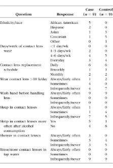
          There were no marked differences in SCL wear behaviors or exposures between case and control subjects. Predominant organisms detected among case and control subjects were Staphylococcus, Propionibacterium, Streptococcus, and Corynebacterium. Marginally higher levels of Neisseria were found in three of nine cases but zero of nine control samples ( P = 0.056). A potentially deleterious missense single nucleotide polymorphism (SNP) variant in IL-6 Signal Transducer ( IL6ST) was found in seven of eight cases and zero of nine controls (rs2228046; P = 0.03). The concentration of tear IL-6 was significantly higher in cases (4.5 [range, 2.1 to 6.2] pg/mL) versus controls (3.5 [range, 2.5 to 6.6] Pg/mL; = 0.02).

          Conclusions

          Tear IL-6 concentration was higher, and SNP variants were detected in subjects with a history of CIEs compared with healthy controls. The synthesis, signaling, and ocular surface cytokine concentration of IL-6 may be related to susceptibility to CIE. A larger study population is required to further explore relationships between genetic variations, the ocular surface microbiome, inflammatory mediators, and environmental exposures.

          Related collections

          Most cited references53

          • Record: found
          • Abstract: found
          • Article: not found

          Interleukin-6 and Diabetes: The Good, the Bad, or the Indifferent?

          Inflammatory mechanisms play a key role in the pathogenesis of type 1 diabetes. Individuals who progress to type 2 diabetes display features of low-grade inflammation years in advance of disease onset. This low-grade inflammation has been proposed to be involved in the pathogenetic processes causing type 2 diabetes. Mediators of inflammation such as tumor necrosis factor-alpha, interleukin (IL)-1beta, the IL-6 family of cytokines, IL-18, and certain chemokines have been proposed to be involved in the events causing both forms of diabetes. IL-6 has in addition to its immunoregulatory actions been proposed to affect glucose homeostasis and metabolism directly and indirectly by action on skeletal muscle cells, adipocytes, hepatocytes, pancreatic beta-cells, and neuroendocrine cells. Here we argue that IL-6 action-in part regulated by variance in the IL-6 and IL-6alpha receptor genes-contributes to, but is probably neither necessary nor sufficient for, the development of both type 1 and type 2 diabetes. Thus, the two types of diabetes are also in this respect less apart than apparent. However, the mechanisms are not clear, and we therefore propose future directions for studies in this field.
            Bookmark
            • Record: found
            • Abstract: found
            • Article: not found

            Diversity of bacteria at healthy human conjunctiva.

            Ocular surface (OS) microbiota contributes to infectious and autoimmune diseases of the eye. Comprehensive analysis of microbial diversity at the OS has been impossible because of the limitations of conventional cultivation techniques. This pilot study aimed to explore true diversity of human OS microbiota using DNA sequencing-based detection and identification of bacteria. Composition of the bacterial community was characterized using deep sequencing of the 16S rRNA gene amplicon libraries generated from total conjunctival swab DNA. The DNA sequences were classified and the diversity parameters measured using bioinformatics software ESPRIT and MOTHUR and tools available through the Ribosomal Database Project-II (RDP-II). Deep sequencing of conjunctival rDNA from four subjects yielded a total of 115,003 quality DNA reads, corresponding to 221 species-level phylotypes per subject. The combined bacterial community classified into 5 phyla and 59 distinct genera. However, 31% of all DNA reads belonged to unclassified or novel bacteria. The intersubject variability of individual OS microbiomes was very significant. Regardless, 12 genera-Pseudomonas, Propionibacterium, Bradyrhizobium, Corynebacterium, Acinetobacter, Brevundimonas, Staphylococci, Aquabacterium, Sphingomonas, Streptococcus, Streptophyta, and Methylobacterium-were ubiquitous among the analyzed cohort and represented the putative "core" of conjunctival microbiota. The other 47 genera accounted for <4% of the classified portion of this microbiome. Unexpectedly, healthy conjunctiva contained many genera that are commonly identified as ocular surface pathogens. The first DNA sequencing-based survey of bacterial population at the conjunctiva have revealed an unexpectedly diverse microbial community. All analyzed samples contained ubiquitous (core) genera that included commensal, environmental, and opportunistic pathogenic bacteria.
              Bookmark
              • Record: found
              • Abstract: found
              • Article: not found

              Characterization of the normal microbiota of the ocular surface.

              The ocular surface is continually exposed to the environment and as a consequence to different types of microbes, but whether there is a normal microbiota of the ocular surface remains unresolved. Using traditional microbial culture techniques has shown that <80% of swabs of the conjunctiva yield cultivable microbes. These usually belong to the bacterial types of the coagulase-negative staphylococci, Propionibacterium sp., with low frequency of isolation of bacteria such as Staphylococcus aureus, Micrococcus sp., Gram-negative bacteria or fungi. Even when these are grown, the numbers of colony forming units (cfu) per swab of the conjunctiva is usually much less than 100 cfu. Swabs of the lid more commonly result in microbial growth, of the same species as from the conjunctiva and slightly higher cfu. Contact lenses have also been cultured, and they yield similar microbial types. Microbes can be isolated from the ocular surface almost immediately after birth. The advent of molecular techniques for microbial identification based on 16S rRNA sequencing has opened up the possibility of determining whether there are non-cultivable microbes that can colonise the ocular surface. Additionally, use of these techniques with cross-sectional and longitudinal studies may help to understand whether the ocular surface harbours its own unique microbiota, or whether the microbiota are only transiently present. Copyright © 2013 Elsevier Ltd. All rights reserved.
                Bookmark

                Author and article information

                Journal
                Invest Ophthalmol Vis Sci
                Invest. Ophthalmol. Vis. Sci
                iovs
                Invest Ophthalmol Vis Sci
                IOVS
                Investigative Ophthalmology & Visual Science
                The Association for Research in Vision and Ophthalmology
                0146-0404
                1552-5783
                December 2018
                : 59
                : 15
                : 5752-5761
                Affiliations
                [1 ]Tufts Medical Center, Center for Translational Ocular Immunology, Boston, Massachusetts, United States
                [2 ]University of New South Wales School of Optometry and Vision Science, Kensington, New South Wales, Australia
                [3 ]Department of Ophthalmology, University of Washington Medical School, Seattle, Washington, United States
                [4 ]Department of Population and Quantitative Health Sciences, Case Western Reserve University, Institute for Computational Biology, Cleveland, Ohio, United States
                [5 ]Johnson & Johnson Vision Care, Inc., Jacksonville, Florida, United States
                [6 ]University of Houston College of Optometry, Houston, Texas, United States
                Author notes
                Correspondence: Kathryn Richdale, University of Houston College of Optometry, 4901 Calhoun Road, Houston, TX 77024, USA; richdale@ 123456uh.edu .
                Article
                iovs-59-13-06 IOVS-18-24845R2
                10.1167/iovs.18-24845
                6281277
                30516819
                22aaf5f9-ec4b-4d49-9e28-bca5ced55708
                Copyright 2018 The Authors

                This work is licensed under a Creative Commons Attribution-NonCommercial-NoDerivatives 4.0 International License.

                History
                : 10 July 2018
                : 15 October 2018
                Categories
                Clinical and Epidemiologic Research

                tear cytokines,ocular microbiome,whole exome sequencing,soft contact lenses,corneal infiltrative events

                Comments

                Comment on this article