7
views
0
recommends
+1 Recommend
0 collections
    0
    shares
      • Record: found
      • Abstract: found
      • Article: found
      Is Open Access

      Loss of mitochondrial enzyme GPT2 causes early neurodegeneration in locus coeruleus

      research-article

      Read this article at

      Bookmark
          There is no author summary for this article yet. Authors can add summaries to their articles on ScienceOpen to make them more accessible to a non-specialist audience.

          Abstract

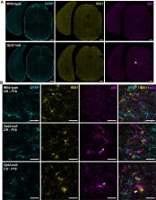
          Locus coeruleus (LC) is among the first brain areas to degenerate in Alzheimer’s disease and Parkinson’s disease; however, the underlying causes for the vulnerability of LC neurons are not well defined. Here we report a novel mechanism of degeneration of LC neurons caused by loss of the mitochondrial enzyme glutamate pyruvate transaminase 2 (GPT2). GPT2 Deficiency is a newly-recognized childhood neurometabolic disorder. The GPT2 enzyme regulates cell growth through replenishment of tricarboxylic acid (TCA) cycle intermediates and modulation of amino acid metabolism. In Gpt2-null mice, we observe an early loss of tyrosine hydroxylase (TH)-positive neurons in LC and reduced soma size at postnatal day 18. Gpt2-null LC shows selective positive Fluoro-Jade C staining. Neuron loss is accompanied by selective, prominent microgliosis and astrogliosis in LC. We observe reduced noradrenergic projections to and norepinephrine levels in hippocampus and spinal cord. Whole cell recordings in Gpt2-null LC slices show reduced soma size and abnormal action potentials with altered firing kinetics. Strikingly, we observe early decreases in phosphorylated S6 in Gpt2-null LC, preceding prominent p62 aggregation, increased LC3B-II to LC3B-I ratio, and neuronal loss. These data are consistent with a possible mechanism involving deficiency in protein synthesis and cell growth, associated subsequently with abnormal autophagy and neurodegeneration. As compared to the few genetic animal models with LC degeneration, loss of LC neurons in Gpt2-null mice is developmentally the earliest. Early neuron loss in LC in a model of human neurometabolic disease provides important clues regarding the metabolic vulnerability of LC and may lead to new therapeutic targets.

          Related collections

          Most cited references91

          • Record: found
          • Abstract: found
          • Article: not found

          An RNA-sequencing transcriptome and splicing database of glia, neurons, and vascular cells of the cerebral cortex.

          The major cell classes of the brain differ in their developmental processes, metabolism, signaling, and function. To better understand the functions and interactions of the cell types that comprise these classes, we acutely purified representative populations of neurons, astrocytes, oligodendrocyte precursor cells, newly formed oligodendrocytes, myelinating oligodendrocytes, microglia, endothelial cells, and pericytes from mouse cerebral cortex. We generated a transcriptome database for these eight cell types by RNA sequencing and used a sensitive algorithm to detect alternative splicing events in each cell type. Bioinformatic analyses identified thousands of new cell type-enriched genes and splicing isoforms that will provide novel markers for cell identification, tools for genetic manipulation, and insights into the biology of the brain. For example, our data provide clues as to how neurons and astrocytes differ in their ability to dynamically regulate glycolytic flux and lactate generation attributable to unique splicing of PKM2, the gene encoding the glycolytic enzyme pyruvate kinase. This dataset will provide a powerful new resource for understanding the development and function of the brain. To ensure the widespread distribution of these datasets, we have created a user-friendly website (http://web.stanford.edu/group/barres_lab/brain_rnaseq.html) that provides a platform for analyzing and comparing transciption and alternative splicing profiles for various cell classes in the brain. Copyright © 2014 the authors 0270-6474/14/3411929-19$15.00/0.
            Bookmark
            • Record: found
            • Abstract: found
            • Article: not found

            LC3, a mammalian homologue of yeast Apg8p, is localized in autophagosome membranes after processing.

            Little is known about the protein constituents of autophagosome membranes in mammalian cells. Here we demonstrate that the rat microtubule-associated protein 1 light chain 3 (LC3), a homologue of Apg8p essential for autophagy in yeast, is associated to the autophagosome membranes after processing. Two forms of LC3, called LC3-I and -II, were produced post-translationally in various cells. LC3-I is cytosolic, whereas LC3-II is membrane bound. The autophagic vacuole fraction prepared from starved rat liver was enriched with LC3-II. Immunoelectron microscopy on LC3 revealed specific labelling of autophagosome membranes in addition to the cytoplasmic labelling. LC3-II was present both inside and outside of autophagosomes. Mutational analyses suggest that LC3-I is formed by the removal of the C-terminal 22 amino acids from newly synthesized LC3, followed by the conversion of a fraction of LC3-I into LC3-II. The amount of LC3-II is correlated with the extent of autophagosome formation. LC3-II is the first mammalian protein identified that specifically associates with autophagosome membranes.
              Bookmark
              • Record: found
              • Abstract: found
              • Article: not found

              A ubiquitin-like system mediates protein lipidation.

              Autophagy is a dynamic membrane phenomenon for bulk protein degradation in the lysosome/vacuole. Apg8/Aut7 is an essential factor for autophagy in yeast. We previously found that the carboxy-terminal arginine of nascent Apg8 is removed by Apg4/Aut2 protease, leaving a glycine residue at the C terminus. Apg8 is then converted to a form (Apg8-X) that is tightly bound to the membrane. Here we report a new mode of protein lipidation. Apg8 is covalently conjugated to phosphatidylethanolamine through an amide bond between the C-terminal glycine and the amino group of phosphatidylethanolamine. This lipidation is mediated by a ubiquitination-like system. Apg8 is a ubiquitin-like protein that is activated by an E1 protein, Apg7 (refs 7, 8), and is transferred subsequently to the E2 enzymes Apg3/Aut1 (ref. 9). Apg7 activates two different ubiquitin-like proteins, Apg12 (ref. 10) and Apg8, and assigns them to specific E2 enzymes, Apg10 (ref. 11) and Apg3, respectively. These reactions are necessary for the formation of Apg8-phosphatidylethanolamine. This lipidation has an essential role in membrane dynamics during autophagy.
                Bookmark

                Author and article information

                Journal
                9500169
                20475
                Neurobiol Dis
                Neurobiol Dis
                Neurobiology of disease
                0969-9961
                1095-953X
                10 November 2022
                15 October 2022
                28 July 2022
                17 November 2022
                : 173
                : 105831
                Affiliations
                [a ]Department of Molecular Biology, Cell Biology and Biochemistry, Brown University, Providence, RI 02912, USA
                [b ]Center for Translational Neuroscience, Carney Institute for Brain Science and Brown Institute for Translational Science, Brown University, Providence, RI 02912, USA
                [c ]Neuroscience Graduate Program, Brown University, Providence, RI 02912, USA
                [d ]Department of Psychiatry and Behavioral Sciences, Stanford University School of Medicine, Stanford, CA 94035, USA
                Author notes

                Author contributions

                Conceptualization O.B. and E.M.M.; O.B. conducted all experiments; Data curation and Visualization O.B.; Experimental Design and Data Analysis O.B, J.A.K. and E.M.M.; Formal analysis and Writing – Original Draft, O.B. and E.M.M.; Writing – Review & Editing, O.B., J.A.K. and E. M.M.; Supervision, Project Administration and Funding Acquisition, E.M.M.

                [* ]Corresponding author at: Brown University, Laboratories for Molecular Medicine, 70 Ship Street, Providence, RI 02912, USA, eric_morrow@ 123456brown.edu (E.M. Morrow).
                Article
                NIHMS1841815
                10.1016/j.nbd.2022.105831
                9669404
                35908744
                220ae538-e771-4502-b0d1-05f942a38900

                This is an open access article under the CC BY-NC-ND license ( http://creativecommons.org/licenses/by-nc-nd/4.0/).

                History
                Categories
                Article

                Neurosciences
                locus coeruleus,gpt2,neurodegeneration,neurometabolism,neurogenetics,selective vulnerability,proteostasis,autophagy

                Comments

                Comment on this article