0
views
0
recommends
+1 Recommend
0 collections
    0
    shares
      • Record: found
      • Abstract: found
      • Article: found
      Is Open Access

      Non-Pulmonary Vein Triggers of Atrial Fibrillation Are Likely to Arise from Low-Voltage Areas in the Left Atrium

      research-article

      Read this article at

      Bookmark
          There is no author summary for this article yet. Authors can add summaries to their articles on ScienceOpen to make them more accessible to a non-specialist audience.

          Abstract

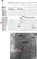
          The pathophysiology of non-pulmonary vein (PV) triggers of atrial fibrillation (AF) is unclear. We hypothesized that left atrial non-PV (LANPV) triggers are associated with atrial tissue degeneration. This study analyzed 431 patients that underwent catheter ablation (mean age 62 yrs, 303 men, 255 paroxysmal AF [pAF] patients). Clinical and electrophysiological characteristics of non-PV trigger were analyzed. Fifty non-PV triggers in 40 patients (9.3%) were documented; LANPV triggers were the most prevalent (n = 19, 38%). LANPV triggers were correlated with non-paroxysmal AF (non-pAF) (OR 3.31, p = 0.04) whereas right atrial non-PV (RANPV) triggers (n = 14) and SVC triggers (n = 17) were not. The voltage at the LANPV sites during SR was 0.3 ± 0.16 mV (p < 0.001 vs. control site). Low-voltage areas (LVAs) in the LA were significantly greater in non-pAF compared to pAF (14.2% vs. 5.8%, p < 0.01). RANPV trigger sites had preserved voltage (0.74 ± 0.48 mV). Long-term outcomes of patients with non-PV triggers treated with tailored targeting strategies were not significantly inferior to those without non-PV triggers. In conclusion, non-PV triggers arise from the LA with degeneration, which may have an important role in AF persistence. A trigger-oriented, patient-tailored ablation strategy considering LA voltage map may be feasible and effective in persistent/recurrent AF.

          Related collections

          Most cited references23

          • Record: found
          • Abstract: found
          • Article: not found

          Tailored atrial substrate modification based on low-voltage areas in catheter ablation of atrial fibrillation.

          Reduced electrogram amplitude has been shown to correlate with diseased myocardium. We describe a novel individualized approach for catheter ablation of atrial fibrillation (AF) based on low-voltage areas (LVAs) in the left atrium (LA). We sought to assess (1) the incidence of LVAs in patients undergoing AF catheter ablation, (2) the distribution of LVAs within the LA, and (3) the effect of an individualized ablation strategy on long-term rhythm outcomes.
            Bookmark
            • Record: found
            • Abstract: found
            • Article: not found

            Catheter ablation of paroxysmal atrial fibrillation initiated by non-pulmonary vein ectopy.

            Most of the ectopic beats initiating paroxysmal atrial fibrillation (PAF) originate from the pulmonary vein (PV). However, only limited data are available on PAF originating from the non-PV areas. Two hundred forty patients with a total of 358 ectopic foci initiating PAF were included. Sixty-eight (28%) patients had AF initiated by ectopic beats (73 foci, 20%) from the non-PV areas, including the left atrial posterior free wall (28, 38.3%), superior vena cava (27, 37.0%), crista terminalis (10, 3.7%), ligament of Marshall (6, 8.2%), coronary sinus ostium (1, 1.4%), and interatrial septum (1, 1.4%). Catheter ablation eliminated AF with acute success rates of 63%, 96%, 100%, 50%, 100%, and 0% in left atrial posterior free wall, superior vena cava, crista terminalis, ligament of Marshall, coronary sinus ostium, and interatrial septum, respectively. During a follow-up period of 22+/-11 months, 43 patients (63.2%) were free of antiarrhythmic drugs without AF recurrence. Ectopic beats initiating PAF can originate from the non-PV areas, and catheter ablation of the non-PV ectopy has a moderate efficacy in treatment of PAF.
              Bookmark
              • Record: found
              • Abstract: found
              • Article: not found

              Molecular determinants of cardiac fibroblast electrical function and therapeutic implications for atrial fibrillation.

              Cardiac fibroblasts account for about 75% of all cardiac cells, but because of their small size contribute only ∼10-15% of total cardiac cell volume. They play a crucial role in cardiac pathophysiology. For a long time, it has been recognized that fibroblasts and related cell types are the principal sources of extracellular matrix (ECM) proteins, which organize cardiac cellular architecture. In disease states, fibroblast production of increased quantities of ECM proteins leads to tissue fibrosis, which can impair both mechanical and electrical function of the heart, contributing to heart failure and arrhythmogenesis. Atrial fibrosis is known to play a particularly important role in atrial fibrillation (AF). This review article focuses on recent advances in understanding the molecular electrophysiology of cardiac fibroblasts. Cardiac fibroblasts express a variety of ion channels, in particular voltage-gated K(+) channels and non-selective cation channels of the transient receptor potential (TRP) family. Both K(+) and TRP channels are important determinants of fibroblast function, with TRP channels acting as Ca(2+)-entry pathways that stimulate fibroblast differentiation into secretory myofibroblast phenotypes producing ECM proteins. Fibroblasts can couple to cardiomyocytes and substantially affect their cellular electrical properties, including conduction, resting potential, repolarization, and excitability. Co-cultured preparations of cardiomyocytes and fibroblasts generate arrhythmias by a variety of mechanisms, including spontaneous impulse formation and rotor-driven reentry. In addition, the excess ECM proteins produced by fibroblasts can interrupt cardiomyocyte-bundle continuity, leading to local conduction disturbances and reentrant arrhythmias. A better understanding of the electrical properties of fibroblasts should lead to an improved comprehension of AF pathophysiology and a variety of novel targets for antiarrhythmic intervention.
                Bookmark

                Author and article information

                Contributors
                y_mukai@junnai.org
                Journal
                Sci Rep
                Sci Rep
                Scientific Reports
                Nature Publishing Group UK (London )
                2045-2322
                22 August 2019
                22 August 2019
                2019
                : 9
                : 12271
                Affiliations
                [1 ]ISNI 0000 0001 2242 4849, GRID grid.177174.3, Department of Cardiovascular Medicine, , Kyushu University Graduate School of Medical Sciences, ; Fukuoka, Japan
                [2 ]ISNI 0000 0001 2242 4849, GRID grid.177174.3, Department of Health Sciences, Graduate School of Medical Sciences, , Kyushu University, ; Fukuoka, Japan
                Article
                48669
                10.1038/s41598-019-48669-1
                6706423
                31439861
                21284855-dba2-4e07-bb7f-b889c0b68ae4
                © The Author(s) 2019

                Open Access This article is licensed under a Creative Commons Attribution 4.0 International License, which permits use, sharing, adaptation, distribution and reproduction in any medium or format, as long as you give appropriate credit to the original author(s) and the source, provide a link to the Creative Commons license, and indicate if changes were made. The images or other third party material in this article are included in the article’s Creative Commons license, unless indicated otherwise in a credit line to the material. If material is not included in the article’s Creative Commons license and your intended use is not permitted by statutory regulation or exceeds the permitted use, you will need to obtain permission directly from the copyright holder. To view a copy of this license, visit http://creativecommons.org/licenses/by/4.0/.

                History
                : 5 March 2019
                : 7 August 2019
                Funding
                Funded by: FundRef https://doi.org/10.13039/100002491, Bristol-Myers Squibb (Bristol-Myers Squibb Company);
                Award ID: #GAKF850098
                Award Recipient :
                Categories
                Article
                Custom metadata
                © The Author(s) 2019

                Uncategorized
                interventional cardiology,outcomes research
                Uncategorized
                interventional cardiology, outcomes research

                Comments

                Comment on this article