7
views
0
recommends
+1 Recommend
0 collections
    0
    shares
      • Record: found
      • Abstract: found
      • Article: found
      Is Open Access

      One Odontogenic Cell-Population Contributes to the Development of the Mouse Incisors and of the Oral Vestibule

      research-article

      Read this article at

      Bookmark
          There is no author summary for this article yet. Authors can add summaries to their articles on ScienceOpen to make them more accessible to a non-specialist audience.

          Abstract

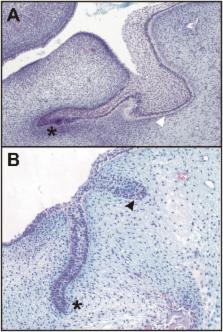
          The area of the oral vestibule is often a place where pathologies appear (e.g., peripheral odontomas). The origin of these pathologies is not fully understood. In the present study, we traced a cell population expressing Sonic hedgehog ( Shh) from the beginning of tooth development using Cre-LoxP system in the lower jaw of wild-type (WT) mice. We focused on Shh expression in the area of the early appearing rudimentary incisor germs located anteriorly to the prospective incisors. The localization of the labelled cells in the incisor germs and also in the inner epithelial layer of the vestibular anlage showed that the first very early developmental events in the lower incisor area are common to the vestibulum oris and the prospective incisor primordia in mice. Scanning electron microscopic analysis of human historical tooth-like structures found in the vestibular area of jaws confirmed their relation to teeth and thus the capability of the vestibular tissue to form teeth. The location of labelled cells descendant of the early appearing Shh expression domain related to the rudimentary incisor anlage not only in the rudimentary and functional incisor germs but also in the externally located anlage of the oral vestibule documented the odontogenic potential of the vestibular epithelium. This potential can be awakened under pathological conditions and become a source of pathologies in the vestibular area.

          Related collections

          Most cited references12

          • Record: found
          • Abstract: found
          • Article: not found

          Sox2+ stem cells contribute to all epithelial lineages of the tooth via Sfrp5+ progenitors.

          The continuously growing mouse incisor serves as a valuable model to study stem cell regulation during organ renewal. Epithelial stem cells are localized in the proximal end of the incisor in the labial cervical loop. Here, we show that the transcription factor Sox2 is a specific marker for these stem cells. Sox2+ cells became restricted to the labial cervical loop during tooth morphogenesis, and they contributed to the renewal of enamel-producing ameloblasts as well as all other epithelial cell lineages of the tooth. The early progeny of Sox2-positive stem cells transiently expressed the Wnt inhibitor Sfrp5. Sox2 expression was regulated by the tooth initiation marker FGF8 and specific miRNAs, suggesting a fine-tuning to maintain homeostasis of the dental epithelium. The identification of Sox2 as a marker for the dental epithelial stem cells will facilitate further studies on their lineage segregation and differentiation during tooth renewal. Copyright © 2012 Elsevier Inc. All rights reserved.
            Bookmark
            • Record: found
            • Abstract: found
            • Article: not found

            Oral manifestations of Ellis-van Creveld syndrome: report of two siblings with unusual dental anomalies.

            Ellis-van Creveld syndrome (EvC), also called chondroectodermal dysplasia, is a rare occurrence inherited as an autosomal recessive disease. Despite the fact that oral manifestations play an important role in the diagnosis criteria for EvC, few detailed reports have been published in the dental literature. This articles presents two siblings with EvC, a boy aged 9 years and a girl aged 7 1/2 years, a product of unaffected first cousin parents. The patients manifests: chondrodysplasia of tubular bones resulting in disproportionate dwarfism, polydactyly and syndactyly of hands and feet, severe dystrophic nails, multiple broad labial frenula with abnormal attachments, congenital missing incisors, anomalous teeth, bilateral partial clefts of the alveolar bone, and malocclusion. Other features noted in either cases are: congenital heart defect, median notch of the upper lip, shovel-shaped incisors and taurodontism. Of the unusual dental findings observed in our patients are talon cusp, reduced crown size, supernumerary tooth, and early eruption of teeth. Because half of the cases with EvC have cardiac malformation, dental treatment must be performed under prophylactic antibiotic coverage. Dentists play an important role in early diagnosis and control of dental problem of this condition.
              Bookmark
              • Record: found
              • Abstract: found
              • Article: not found

              Expression of the stem cell marker, SOX2, in ameloblastoma and dental epithelium.

              Ameloblastomas are locally invasive odontogenic tumors that exhibit a high rate of recurrence and often associate with the third molars. They are suggested to originate from dental epithelium because the tumor cells resemble epithelial cells of developing teeth. Expression of the transcription factor SOX2 has been previously localized in epithelial stem and progenitor cells in developing teeth as well as in various tumors. Here, we show that SOX2 is expressed in the epithelial cells of follicular and plexiform ameloblastomas. SOX2 was localized in the dental lamina of developing human primary molars. It was also expressed in the fragmented dental lamina associated with the third molars and in the epithelium budding from its posterior aspect in mice. However, no SOX2 expression was detected in either Hertwig's epithelial root sheath directing the formation of roots or in the epithelial cell rests of Malassez covering the completed roots. SOX2 was associated with supernumerary tooth formation in odontoma-like tumors induced by Wnt signal activation in mice. We propose that SOX2 functions in maintaining the progenitor state of epithelium in ameloblastomas and that ameloblastomas may originate from SOX2-expressing dental lamina epithelium.
                Bookmark

                Author and article information

                Contributors
                Role: Editor
                Journal
                PLoS One
                PLoS ONE
                plos
                plosone
                PLoS ONE
                Public Library of Science (San Francisco, CA USA )
                1932-6203
                9 September 2016
                2016
                : 11
                : 9
                : e0162523
                Affiliations
                [1 ]Department of Teratology, Institute of Experimental Medicine, Academy of Sciences of the Czech Republic, 14220, Prague 4, Czech Republic
                [2 ]Department of Anthropology and Human Genetics, Faculty of Science, Charles University, Prague, Czech Republic
                [3 ]Department of Anthropology, Faculty of Natural Sciences, Comenius University in Bratislava, Bratislava, Slovak Republic
                [4 ]Department of Cell Biology, Faculty of Science, Charles University, Prague, Czech Republic
                University of Texas at Austin Dell Medical School, UNITED STATES
                Author notes

                Competing Interests: The authors declare that they have no competing interests.

                • Conceptualization: MH.

                • Funding acquisition: MH SB.

                • Investigation: MH OZ SB KL KDT MD LHS.

                • Methodology: MH SB.

                • Project administration: MH SB.

                • Resources: MH SB.

                • Validation: MH OZ SB.

                • Visualization: MH OZ SB KL KDT MD LHS.

                • Writing – original draft: MH SB.

                • Writing – review & editing: MH OZ SB KL KDT MD LHS.

                Article
                PONE-D-16-17851
                10.1371/journal.pone.0162523
                5017683
                27611193
                20e9743f-ec88-4637-8413-13667ffa5f98
                © 2016 Hovorakova et al

                This is an open access article distributed under the terms of the Creative Commons Attribution License, which permits unrestricted use, distribution, and reproduction in any medium, provided the original author and source are credited.

                History
                : 10 May 2016
                : 24 August 2016
                Page count
                Figures: 5, Tables: 2, Pages: 16
                Funding
                Funded by: funder-id http://dx.doi.org/10.13039/501100001824, Grantová Agentura eské Republiky;
                Award ID: P305/12/1766
                Award Recipient :
                Funded by: funder-id http://dx.doi.org/10.13039/501100003194, Agentúra Ministerstva kolstva, vedy, výskumu a portu SR;
                Award ID: VEGA 1/0442/13
                Award Recipient :
                Funded by MH: Grant Agency of the Czech Republic (P305/12/1766) and SB: Ministry of Education, Science, Research and Sport of the Slovak Republic (VEGA 1/0442/13).
                Categories
                Research Article
                Biology and Life Sciences
                Anatomy
                Digestive System
                Teeth
                Incisors
                Medicine and Health Sciences
                Anatomy
                Digestive System
                Teeth
                Incisors
                Biology and Life Sciences
                Anatomy
                Head
                Jaw
                Teeth
                Incisors
                Medicine and Health Sciences
                Anatomy
                Head
                Jaw
                Teeth
                Incisors
                Biology and Life Sciences
                Physiology
                Digestive Physiology
                Dentition
                Medicine and Health Sciences
                Physiology
                Digestive Physiology
                Dentition
                Biology and Life Sciences
                Anatomy
                Biological Tissue
                Epithelium
                Medicine and Health Sciences
                Anatomy
                Biological Tissue
                Epithelium
                Biology and Life Sciences
                Developmental Biology
                Embryology
                Embryos
                Biology and Life Sciences
                Anatomy
                Head
                Jaw
                Medicine and Health Sciences
                Anatomy
                Head
                Jaw
                Research and Analysis Methods
                Model Organisms
                Animal Models
                Mouse Models
                Research and Analysis Methods
                Microscopy
                Electron Microscopy
                Scanning Electron Microscopy
                Biology and Life Sciences
                Organisms
                Animals
                Vertebrates
                Amniotes
                Reptiles
                Custom metadata
                All relevant data are within the paper and its Supporting Information files.

                Uncategorized
                Uncategorized

                Comments

                Comment on this article