27
views
0
recommends
+1 Recommend
0 collections
    0
    shares
      • Record: found
      • Abstract: found
      • Article: not found

      Glycogen in the uterus and fallopian tubes is an important source of glucose during early pregnancy†

      1
      Biology of Reproduction
      Oxford University Press (OUP)

      Read this article at

      ScienceOpenPublisherPubMed
      Bookmark
          There is no author summary for this article yet. Authors can add summaries to their articles on ScienceOpen to make them more accessible to a non-specialist audience.

          Abstract

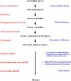
          Pregnancy loss is common during the peri-implantation period in mammals when glucose is required for both embryonic development and decidualization of the endometrium. As the uterus cannot synthesize glucose, all glucose must come directly from maternal circulation as needed or transiently stored as the macromolecule glycogen. Glycogen acts as a glucose reservoir, storing up to 55 000 glucose moieties per molecule. Endometrial glycogen concentrations are correlated with fertility in humans, indicating that glycogen is an essential source of glucose during early pregnancy. In humans and primates, endometrial glycogen concentrations peak during the luteal phase due to progesterone. In contrast, in rats and mink, estradiol triggers an accumulation of uterine glycogen during proestrus and estrus. In mated rats, the glycogen content of the endometrium increases again after implantation due to high levels of glycogen stored in the decidua. In mink, endometrial glycogen reserves are localized in the uterine epithelia at estrus. These reserves are mobilized before implantation, suggesting they are used to support embryonic growth. Uterine glycogen concentrations continue to decrease after implantation in mink, probably due to a lack of decidualization. How ovarian steroids stimulate glycogenesis in the endometrium is unclear, but current evidence suggests that estradiol/progesterone interacts with insulin or insulin-like growth factor signaling. In summary, endometrial glycogen is an essential source of glucose during the peri-implantation period. More work is needed to characterize differences among species, elucidate the fate of the glucose liberated from glycogen, and understand how ovarian steroids regulate glycogen metabolism in the uterus.

          Related collections

          Most cited references101

          • Record: found
          • Abstract: found
          • Article: found
          Is Open Access

          Glycogen metabolism in humans☆☆☆

          In the human body, glycogen is a branched polymer of glucose stored mainly in the liver and the skeletal muscle that supplies glucose to the blood stream during fasting periods and to the muscle cells during muscle contraction. Glycogen has been identified in other tissues such as brain, heart, kidney, adipose tissue, and erythrocytes, but glycogen function in these tissues is mostly unknown. Glycogen synthesis requires a series of reactions that include glucose entrance into the cell through transporters, phosphorylation of glucose to glucose 6-phosphate, isomerization to glucose 1-phosphate, and formation of uridine 5ʹ-diphosphate-glucose, which is the direct glucose donor for glycogen synthesis. Glycogenin catalyzes the formation of a short glucose polymer that is extended by the action of glycogen synthase. Glycogen branching enzyme introduces branch points in the glycogen particle at even intervals. Laforin and malin are proteins involved in glycogen assembly but their specific function remains elusive in humans. Glycogen is accumulated in the liver primarily during the postprandial period and in the skeletal muscle predominantly after exercise. In the cytosol, glycogen breakdown or glycogenolysis is carried out by two enzymes, glycogen phosphorylase which releases glucose 1-phosphate from the linear chains of glycogen, and glycogen debranching enzyme which untangles the branch points. In the lysosomes, glycogen degradation is catalyzed by α-glucosidase. The glucose 6-phosphatase system catalyzes the dephosphorylation of glucose 6-phosphate to glucose, a necessary step for free glucose to leave the cell. Mutations in the genes encoding the enzymes involved in glycogen metabolism cause glycogen storage diseases.
            Bookmark
            • Record: found
            • Abstract: found
            • Article: not found

            Conception, early pregnancy loss, and time to clinical pregnancy: a population-based prospective study.

            To examine rates of conception and pregnancy loss and their relations with time to clinical pregnancy and reproductive outcomes. A prospective observational study. Population-based cohort in China. Five hundred eighteen healthy newly married women who intended to conceive. Upon stopping contraception, daily records of vaginal bleeding and daily first-morning urine specimens were obtained for < or =1 year or until a clinical pregnancy was achieved. Daily urinary hCG was assayed to detect early pregnancy loss (EPL). None. Conception, pregnancy loss, and time to clinical pregnancy. The conception rate per cycle was 40% over the first 12 months. Of the 618 detectable conceptions, 49 (7.9%) ended in clinical spontaneous abortion, and 152 (24.6%) in EPL. Early pregnancy loss was detected in 14% of all the cycles without clinically recognized pregnancy, but the frequencies were lower among women with delayed time to clinical pregnancy. Early pregnancy loss in the preceding cycle was associated with increased odds of conception (odds ratio [OR], 2.6; 95% confidence interval [CI], 1.8-3.9), clinical pregnancy (OR, 2.0; 95% CI, 1.3-3.0), and EPL (OR, 2.4; 95% CI, 1.4-4.2) but was not associated with spontaneous abortion, low birth weight, or preterm birth in the subsequent cycle. We demonstrated substantial EPL in the non-clinically pregnant cycles and a positive relation between EPL and subsequent fertility.
              Bookmark
              • Record: found
              • Abstract: found
              • Article: not found

              Uterine glands provide histiotrophic nutrition for the human fetus during the first trimester of pregnancy.

              Providing adequate nutrition to the fetus is key to a successful pregnancy. The interstitial form of implantation displayed by the human blastocyst is generally associated with early onset of maternal blood flow to the developing placenta, and hence hemotrophic exchange. However, the recent finding that the maternal intraplacental circulation is not fully established until the third month of gestation suggests that human fetal nutrition may be initially histiotrophic. We therefore investigated activity of the uterine glands during early pregnancy. We demonstrate here that these glands remain active until at least wk 10 of pregnancy, and that their secretions are delivered freely into the placental intervillous space. We also demonstrate phagocytic uptake by the placental syncytiotrophoblast of two glycoproteins, the mucin MUC-1 and glycodelin A, synthesized in the maternal glands. Glycodelin was also detected within the epithelium of the secondary yolk sac lining the exocoelomic cavity, indicating that the yolk sac may play an important role in nutrient exchange before vascularisation of the chorionic villi. Our findings demonstrate that the uterine glands are an important source of nutrients during organogenesis, when metabolism is essentially anaerobic.
                Bookmark

                Author and article information

                Journal
                Biology of Reproduction
                Oxford University Press (OUP)
                0006-3363
                1529-7268
                August 2019
                August 01 2019
                June 14 2019
                August 2019
                August 01 2019
                June 14 2019
                : 101
                : 2
                : 297-305
                Affiliations
                [1 ]Department of Animal Science, University of Illinois at Urbana-Champaign, Urbana, Illinois, USA
                Article
                10.1093/biolre/ioz102
                31201425
                1f2d9571-3236-4ba7-9e21-74bb9d286143
                © 2019

                https://academic.oup.com/journals/pages/open_access/funder_policies/chorus/standard_publication_model

                History

                Comments

                Comment on this article