5
views
0
recommends
+1 Recommend
0 collections
    0
    shares
      • Record: found
      • Abstract: found
      • Article: found
      Is Open Access

      Case report and literature review: Fabry disease misdiagnosing as polymyalgia rheumatica

      research-article

      Read this article at

      Bookmark
          There is no author summary for this article yet. Authors can add summaries to their articles on ScienceOpen to make them more accessible to a non-specialist audience.

          Abstract

          Rationale:

          The clinical manifestations of Fabry disease affect the nerves, kidneys, heart, skin, gastrointestinal tract and eyes. Our aim is to familiarize people with the FD diagnostic process by reporting this case.

          Patient concerns:

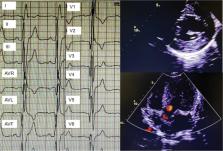
          A 79-year-old-male patient presented with muscle pain and weakness in the extremities, also with an increasing erythrocyte sedimentation rate and C-reactive protein. Further examinations revealed that multiple organ involvement, such as rash, myocardial hypertrophy, peripheral neuropathy.

          Diagnoses:

          Cardiac MR demonstrated hypertrophic cardiomyopathy, myocardial fibrosis and low myocardial T1 value. The patient was eventually diagnosed with Fabry disease through proteomics and genetic testing.

          Interventions:

          The treatment is enzyme replacement therapy (ERT). But this patient could not afford ERT and was given only general symptomatic treatment, pregabalin, and a gradual reduction in glucocorticoid.

          Outcomes:

          The patient’s symptoms of joint pain and muscle weakness reduced significantly, and ESR and CRP had decreased to normal.

          Lessons:

          FD is a rare disease and difficult to diagnose, but rare does not mean invisible. FD may present with signs and symptoms of rheumatic diseases. Rheumatologists should be aware and concerned about this disease.

          Related collections

          Most cited references19

          • Record: found
          • Abstract: found
          • Article: not found
          Is Open Access

          Fabry disease revisited: Management and treatment recommendations for adult patients.

          Fabry disease is an X-linked lysosomal storage disorder caused by mutations in the GLA gene leading to deficient α-galactosidase A activity, glycosphingolipid accumulation, and life-threatening complications. Phenotypes vary from the "classic" phenotype, with pediatric onset and multi-organ involvement, to later-onset, a predominantly cardiac phenotype. Manifestations are diverse in female patients in part due to variations in residual enzyme activity and X chromosome inactivation patterns. Enzyme replacement therapy (ERT) and adjunctive treatments can provide significant clinical benefit. However, much of the current literature reports outcomes after late initiation of ERT, once substantial organ damage has already occurred. Updated monitoring and treatment guidelines for pediatric patients with Fabry disease have recently been published. Expert physician panels were convened to develop updated, specific guidelines for adult patients. Management of adult patients depends on 1) a personalized approach to care, reflecting the natural history of the specific disease phenotype; 2) comprehensive evaluation of disease involvement prior to ERT initiation; 3) early ERT initiation; 4) thorough routine monitoring for evidence of organ involvement in non-classic asymptomatic patients and response to therapy in treated patients; 5) use of adjuvant treatments for specific disease manifestations; and 6) management by an experienced multidisciplinary team.
            Bookmark
            • Record: found
            • Abstract: found
            • Article: not found

            Fabry's disease.

            Fabry's disease is an X-linked lysosomal storage disorder caused by abnormalities in the GLA gene, which leads to a deficiency in alpha-galactosidase A. The consequent abnormal accumulation of glycosphingolipids results in several clinical signs and symptoms and substantial morbidity and mortality. This review covers all basic aspects of the disease such as epidemiology, pathophysiology, clinical presentation by systems, diagnosis, management, prevention, and repercussions on quality of life. With the development of enzyme replacement therapy in the past few years, early initiation of treatment is key for improvement in major affected organs with decreased cardiac mass and stabilisation of kidney function, and improvement in neuropathic pain, sweating, gastrointestinal symptoms, hearing loss, and pulmonary symptoms. However, treatment of individual symptoms in addition to enzyme replacement therapy seems to be needed for many patients, especially those who have had some degree of organ dysfunction. Additional data are needed to document long-term treatment outcomes.
              Bookmark
              • Record: found
              • Abstract: found
              • Article: found

              Noncontrast T1 mapping for the diagnosis of cardiac amyloidosis.

              This study sought to explore the potential role of noncontrast myocardial T1 mapping for detection of cardiac involvement in patients with primary amyloid light-chain (AL) amyloidosis. Cardiac involvement carries a poor prognosis in systemic AL amyloidosis. Late gadolinium enhancement (LGE) cardiac magnetic resonance (CMR) is useful for the detection of cardiac amyloid, but characteristic LGE patterns do not always occur or they appear late in the disease. Noncontrast characterization of amyloidotic myocardium with T1 mapping may improve disease detection. Furthermore, quantitative assessment of myocardial amyloid load would be of great value. Fifty-three AL amyloidosis patients (14 with no cardiac involvement, 11 with possible involvement, and 28 with definite cardiac involvement based on standard biomarker and echocardiographic criteria) underwent CMR (1.5-T) including noncontrast T1 mapping (shortened modified look-locker inversion recovery [ShMOLLI] sequence) and LGE imaging. These were compared with 36 healthy volunteers and 17 patients with aortic stenosis and a comparable degree of left ventricular hypertrophy as the cardiac amyloid patients. Myocardial T1 was significantly elevated in cardiac AL amyloidosis patients (1,140 ± 61 ms) compared to normal subjects (958 ± 20 ms, p < 0.001) and patients with aortic stenosis (979 ± 51 ms, p < 0.001). Myocardial T1 was increased in AL amyloid even when cardiac involvement was uncertain (1,048 ± 48 ms) or thought absent (1,009 ± 31 ms). A noncontrast myocardial T1 cutoff of 1,020 ms yielded 92% accuracy for identifying amyloid patients with possible or definite cardiac involvement. In the AL amyloidosis cohort, there were significant correlations between myocardial T1 time and indices of systolic and diastolic dysfunction. Noncontrast T1 mapping has high diagnostic accuracy for detecting cardiac AL amyloidosis, correlates well with markers of systolic and diastolic dysfunction, and is potentially more sensitive for detecting early disease than LGE imaging. Elevated myocardial T1 may represent a direct marker of cardiac amyloid load. Further studies are needed to assess the prognostic significance of T1 elevation. Copyright © 2013 American College of Cardiology Foundation. Published by Elsevier Inc. All rights reserved.
                Bookmark

                Author and article information

                Contributors
                Journal
                Medicine (Baltimore)
                Medicine (Baltimore)
                MD
                Medicine
                Lippincott Williams & Wilkins (Hagerstown, MD )
                0025-7974
                1536-5964
                3 November 2023
                03 November 2023
                : 102
                : 44
                : e34630
                Affiliations
                [a ] Shengli Clinical Medical College of Fujian Medical University, Department of Rheumatology and Immunology of Fujian Provincial Hospital, Fuzhou, Fujian, China
                [b ] Shengli Clinical Medical College of Fujian Medical University, Department of Imaging of Fujian Provincial Hospital, Fuzhou, Fujian, China.
                Author notes
                [* ]Correspondence: Gao Fei, Shengli Clinical Medical College of Fujian Medical University, Department of Rheumatology and Immunology of Fujian Provincial Hospital, Fuzhou, Fujian 350001, China (e-mail: feifei7316749@ 123456aliyun.com ).
                Author information
                https://orcid.org/0000-0002-1004-4977
                Article
                00086
                10.1097/MD.0000000000034630
                10627660
                1e1f92d8-fd02-4504-9302-593da9f413b8
                Copyright © 2023 the Author(s). Published by Wolters Kluwer Health, Inc.

                This is an open access article distributed under the Creative Commons Attribution License 4.0 (CCBY), which permits unrestricted use, distribution, and reproduction in any medium, provided the original work is properly cited.

                History
                : 14 May 2023
                : 17 July 2023
                Categories
                5100
                Research Article
                Clinical Case Report
                Custom metadata
                TRUE

                fabry disease,misdiagnosis,multiple organs,polymyalgia rheumatica,rare disease

                Comments

                Comment on this article