2
views
0
recommends
+1 Recommend
0 collections
    0
    shares
      • Record: found
      • Abstract: found
      • Article: found
      Is Open Access

      A predictive model for hip abductor strength and knee extensor strength 12 months after total hip arthroplasty with an interaction term

      research-article

      Read this article at

      Bookmark
          There is no author summary for this article yet. Authors can add summaries to their articles on ScienceOpen to make them more accessible to a non-specialist audience.

          Abstract

          Background

          Identifying populations with poor muscle recovery after total hip arthroplasty (THA) is important for postoperative physical therapy. Preoperative muscle strength is a strong factor that determines postoperative muscle strength. However, this effect may depend on other factors. Thus, predictive models with interaction terms are important for accurately predicting postoperative muscle strength. This study aimed to develop a predictive model for lower muscle strength 12 months after THA which incorporates interaction terms.

          Methods

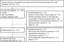
          Subjects were female patients with hip osteoarthritis who underwent unilateral THA. Patients with locomotor disorders, neurological disorders, or postoperative complications were excluded. Hip abductor and knee extensor strength were measured, and a generalized linear model approach with preoperative muscle strength, age, body weight, height, disease duration, physical activity, and leg extension as explanatory variables was used to identify factors that determine muscle strength 12 months after THA. Models with interaction terms between preoperative muscle strength and other explanatory variables were also examined.

          Results

          A total of 82 patients were analyzed. Preoperative muscle strength, age, body weight, physical activity, and disease duration were extracted as factors that significantly and independently determine hip abductor and knee extensor strength. The interaction term between preoperative muscle strength and age was identified as a factor that significantly determines knee extensor strength. Regression coefficients for preoperative knee extensor strength and postoperative muscle strength were significant when age was +1 SD, but not when age was -1 SD.

          Conclusions

          The predictive model demonstrated that lower muscle strength 12 months after THA is determined by preoperative muscle strength, age, weight, physical activity, disease duration, and preoperative muscle strength, with the effect of preoperative muscle strength on knee extensor strength being dependent on age. When predicting postoperative knee extensor strength using preoperative muscle strength, it is important to consider the effect of age.

          Related collections

          Most cited references39

          • Record: found
          • Abstract: found
          • Article: not found

          Skeletal muscle mass and distribution in 468 men and women aged 18-88 yr.

          We employed a whole body magnetic resonance imaging protocol to examine the influence of age, gender, body weight, and height on skeletal muscle (SM) mass and distribution in a large and heterogeneous sample of 468 men and women. Men had significantly (P < 0.001) more SM in comparison to women in both absolute terms (33.0 vs. 21.0 kg) and relative to body mass (38.4 vs. 30.6%). The gender differences were greater in the upper (40%) than lower (33%) body (P < 0.01). We observed a reduction in relative SM mass starting in the third decade; however, a noticeable decrease in absolute SM mass was not observed until the end of the fifth decade. This decrease was primarily attributed to a decrease in lower body SM. Weight and height explained approximately 50% of the variance in SM mass in men and women. Although a linear relationship existed between SM and height, the relationship between SM and body weight was curvilinear because the contribution of SM to weight gain decreased with increasing body weight. These findings indicate that men have more SM than women and that these gender differences are greater in the upper body. Independent of gender, aging is associated with a decrease in SM mass that is explained, in large measure, by a decrease in lower body SM occurring after the fifth decade.
            Bookmark
            • Record: found
            • Abstract: found
            • Article: not found

            Sarcopenia: Aging-Related Loss of Muscle Mass and Function

            Sarcopenia is a loss of muscle mass and function in the elderly that reduces mobility, diminishes quality of life, and can lead to fall-related injuries, which require costly hospitalization and extended rehabilitation. This review focuses on the aging-related structural changes and mechanisms at cellular and subcellular levels underlying changes in the individual motor unit: specifically, the perikaryon of the α-motoneuron, its neuromuscular junction(s), and the muscle fibers that it innervates. Loss of muscle mass with aging, which is largely due to the progressive loss of motoneurons, is associated with reduced muscle fiber number and size. Muscle function progressively declines because motoneuron loss is not adequately compensated by reinnervation of muscle fibers by the remaining motoneurons. At the intracellular level, key factors are qualitative changes in posttranslational modifications of muscle proteins and the loss of coordinated control between contractile, mitochondrial, and sarcoplasmic reticulum protein expression. Quantitative and qualitative changes in skeletal muscle during the process of aging also have been implicated in the pathogenesis of acquired and hereditary neuromuscular disorders. In experimental models, specific intervention strategies have shown encouraging results on limiting deterioration of motor unit structure and function under conditions of impaired innervation. Translated to the clinic, if these or similar interventions, by saving muscle and improving mobility, could help alleviate sarcopenia in the elderly, there would be both great humanitarian benefits and large cost savings for health care systems.
              Bookmark
              • Record: found
              • Abstract: found
              • Article: not found

              Accelerometer data reduction: a comparison of four reduction algorithms on select outcome variables.

              Accelerometers are recognized as a valid and objective tool to assess free-living physical activity. Despite the widespread use of accelerometers, there is no standardized way to process and summarize data from them, which limits our ability to compare results across studies. This paper a) reviews decision rules researchers have used in the past, b) compares the impact of using different decision rules on a common data set, and c) identifies issues to consider for accelerometer data reduction. The methods sections of studies published in 2003 and 2004 were reviewed to determine what decision rules previous researchers have used to identify wearing period, minimal wear requirement for a valid day, spurious data, number of days used to calculate the outcome variables, and extract bouts of moderate to vigorous physical activity (MVPA). For this study, four data reduction algorithms that employ different decision rules were used to analyze the same data set. The review showed that among studies that reported their decision rules, much variability was observed. Overall, the analyses suggested that using different algorithms impacted several important outcome variables. The most stringent algorithm yielded significantly lower wearing time, the lowest activity counts per minute and counts per day, and fewer minutes of MVPA per day. An exploratory sensitivity analysis revealed that the most stringent inclusion criterion had an impact on sample size and wearing time, which in turn affected many outcome variables. These findings suggest that the decision rules employed to process accelerometer data have a significant impact on important outcome variables. Until guidelines are developed, it will remain difficult to compare findings across studies.
                Bookmark

                Author and article information

                Contributors
                dm17017@st.kitasato-u.ac.jp
                takahira@med.kitasato-u.ac.jp
                do-your-best.g@outlook.com
                hw@ahs.kitasato-u.ac.jp
                weardebris@AOL.com
                konsai@tmtv.ne.jp
                Journal
                BMC Musculoskelet Disord
                BMC Musculoskelet Disord
                BMC Musculoskeletal Disorders
                BioMed Central (London )
                1471-2474
                27 September 2021
                27 September 2021
                2021
                : 22
                : 827
                Affiliations
                [1 ]GRID grid.410786.c, ISNI 0000 0000 9206 2938, Graduate School of Medical Sciences, , Kitasato University, ; 1-15-1 Kitasato, Minami-ku, Sagamihara-shi, Kanagawa 252-0373 Japan
                [2 ]Department of Rehabilitation, Zama General Hospital, 1-50-1 Soubudai, Zama-shi, Kanagawa 252-0011 Japan
                [3 ]GRID grid.410786.c, ISNI 0000 0000 9206 2938, Department of Orthopaedic Surgery, , Kitasato University Graduate School of Medical Sciences, ; 1-15-1 Kitasato, Minami-ku, Sagamihara-shi, Kanagawa 252-0373 Japan
                [4 ]GRID grid.410786.c, ISNI 0000 0000 9206 2938, Department of Rehabilitation, , School of Allied Health Sciences, Kitasato University, ; 1-15-1 Kitasato, Minami-ku, Sagamihara-shi, Kanagawa 252-0373 Japan
                [5 ]Institute of Joint Replacement and Rheumatology, Zama General Hospital, 1-50-1 Soubudai, Zama-shi, Kanagawa 252-0011 Japan
                Article
                4719
                10.1186/s12891-021-04719-2
                8474772
                34579703
                1de54ed3-f7b9-46aa-a738-5f4f976de27c
                © The Author(s) 2021

                Open AccessThis article is licensed under a Creative Commons Attribution 4.0 International License, which permits use, sharing, adaptation, distribution and reproduction in any medium or format, as long as you give appropriate credit to the original author(s) and the source, provide a link to the Creative Commons licence, and indicate if changes were made. The images or other third party material in this article are included in the article's Creative Commons licence, unless indicated otherwise in a credit line to the material. If material is not included in the article's Creative Commons licence and your intended use is not permitted by statutory regulation or exceeds the permitted use, you will need to obtain permission directly from the copyright holder. To view a copy of this licence, visit http://creativecommons.org/licenses/by/4.0/. The Creative Commons Public Domain Dedication waiver ( http://creativecommons.org/publicdomain/zero/1.0/) applies to the data made available in this article, unless otherwise stated in a credit line to the data.

                History
                : 19 April 2021
                : 11 September 2021
                Categories
                Research
                Custom metadata
                © The Author(s) 2021

                Orthopedics
                Orthopedics

                Comments

                Comment on this article