24
views
0
recommends
+1 Recommend
0 collections
    0
    shares
      • Record: found
      • Abstract: found
      • Article: found
      Is Open Access

      A Combined In Vitro Imaging and Multi-Scale Modeling System for Studying the Role of Cell Matrix Interactions in Cutaneous Wound Healing

      research-article
      , , *
      PLoS ONE
      Public Library of Science

      Read this article at

      Bookmark
          There is no author summary for this article yet. Authors can add summaries to their articles on ScienceOpen to make them more accessible to a non-specialist audience.

          Abstract

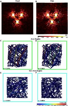
          Many cell types remodel the extracellular matrix of the tissues they inhabit in response to a wide range of environmental stimuli, including mechanical cues. Such is the case in dermal wound healing, where fibroblast migrate into and remodel the provisional fibrin matrix in a complex manner that depends in part on the local mechanical environment and the evolving multi-scale mechanical interactions of the system. In this study, we report on the development of an image-based multi-scale mechanical model that predicts the short-term (24 hours), structural reorganization of a fibrin gel by fibroblasts. These predictive models are based on an in vitro experimental system where clusters of fibroblasts (i.e., explants) were spatially arranged into a triangular geometry onto the surface of fibrin gels that were subjected to either Fixed or Free in-plane mechanical constraints. Experimentally, regional differences in short-term structural remodeling and cell migration were observed for the two gel boundary conditions. A pilot experiment indicated that these small differences in the short-term remodeling of the fibrin gel translate into substantial differences in long-term (4 weeks) remodeling, particularly in terms of collagen production. The multi-scale models were able to predict some regional differences in remodeling and qualitatively similar reorganization patterns for the two boundary conditions. However, other aspects of the model, such as the magnitudes and rates of deformation of gel, did not match the experiments. These discrepancies between model and experiment provide fertile ground for challenging model assumptions and devising new experiments to enhance our understanding of how this multi-scale system functions. These efforts will ultimately improve the predictions of the remodeling process, particularly as it relates to dermal wound healing and the reduction of patient scarring. Such models could be used to recommend patient-specific mechanical-based treatment dependent on parameters such as wound geometry, location, age, and health.

          Related collections

          Most cited references60

          • Record: found
          • Abstract: found
          • Article: not found

          Substrate compliance versus ligand density in cell on gel responses.

          Substrate stiffness is emerging as an important physical factor in the response of many cell types. In agreement with findings on other anchorage-dependent cell lineages, aortic smooth muscle cells are found to spread and organize their cytoskeleton and focal adhesions much more so on "rigid" glass or "stiff" gels than on "soft" gels. Whereas these cells generally show maximal spreading on intermediate collagen densities, the limited spreading on soft gels is surprisingly insensitive to adhesive ligand density. Bell-shaped cell spreading curves encompassing all substrates are modeled by simple functions that couple ligand density to substrate stiffness. Although smooth muscle cells spread minimally on soft gels regardless of collagen, GFP-actin gives a slight overexpression of total actin that can override the soft gel response and drive spreading; GFP and GFP-paxillin do not have the same effect. The GFP-actin cells invariably show an organized filamentous cytoskeleton and clearly indicate that the cytoskeleton is at least one structural node in a signaling network that can override spreading limits typically dictated by soft gels. Based on such results, we hypothesize a central structural role for the cytoskeleton in driving the membrane outward during spreading whereas adhesion reinforces the spreading.
            Bookmark
            • Record: found
            • Abstract: found
            • Article: not found

            The myofibroblast: paradigm for a mechanically active cell.

            Boris Hinz (2010)
            Tissues lose mechanical integrity when our body is injured. To rapidly restore mechanical stability a multitude of cell types can jump into action by acquiring a reparative phenotype-the myofibroblast. Here, I review the known biomechanics of myofibroblast differentiation and action and speculate on underlying mechanisms. Hallmarks of the myofibroblast are secretion of extracellular matrix, development of adhesion structures with the substrate, and formation of contractile bundles composed of actin and myosin. These cytoskeletal features not only enable the myofibroblast to remodel and contract the extracellular matrix but to adapt its activity to changes in the mechanical microenvironment. Rapid repair comes at the cost of tissue contracture due to the inability of the myofibroblast to regenerate tissue. If contracture and ECM remodeling become progressive and manifests as organ fibrosis, the outcome of myofibroblast activity will have more severe consequences than the initial damage. Whereas the pathological consequences of myofibroblast occurrence are of great interest for physicians, their mechano-responsive features render them attractive for physicists and bioengineers. Their well developed cytoskeleton and responsiveness to a plethora of cytokines fascinate cell biologists and biochemists. Finally, the question of the myofibroblast origin intrigues stem cell biologists and developmental biologists-what else can you ask from a truly interdisciplinary cell? Copyright 2009 Elsevier Ltd. All rights reserved.
              Bookmark
              • Record: found
              • Abstract: found
              • Article: not found

              Both Rb/p16INK4a inactivation and telomerase activity are required to immortalize human epithelial cells.

              Normal human cells undergo a limited number of divisions in culture and enter a non-dividing state called replicative senescence. Senescence is accompanied by several changes, including an increase in inhibitors of cyclin-dependent kinases and telomere shortening. The mechanisms by which viral oncogenes reverse these processes are not fully understood, although a general requirement for oncoproteins such as human papillomavirus E6 and E7 has suggested that the p53 and Rb pathways are targeted. Expression of the catalytic component of telomerase, hTERT, alone significantly extends the lifespan of human fibroblasts. Here we show that telomerase activity is not sufficient for immortalization of human keratinocyte or mammary epithelial cells: we find that neither addition of hTERT nor induction of telomerase activity by E6, both of which are active in maintaining telomere length, results in immortalization. Inactivation of the Rb/p16 pathway by E7 or downregulation of p16 expression, in combination with telomerase activity, however, is able to immortalize epithelial cells efficiently. Elimination of p53 and of the DNA-damage-induced G1 checkpoint is not necessary for immortalization, neither is elimination of p19ARF.
                Bookmark

                Author and article information

                Contributors
                Role: Editor
                Journal
                PLoS One
                PLoS ONE
                plos
                plosone
                PLoS ONE
                Public Library of Science (San Francisco, CA USA )
                1932-6203
                3 February 2016
                2016
                : 11
                : 2
                : e0148254
                Affiliations
                [001]Department of Biomedical Engineering, University of Iowa, Iowa City, IA, United States of America
                Université de Technologie de Compiègne, FRANCE
                Author notes

                Competing Interests: The authors have declared that no competing interests exist.

                Conceived and designed the experiments: AD ES. Performed the experiments: AD MA. Analyzed the data: AD MA ES. Contributed reagents/materials/analysis tools: AD MA ES. Wrote the paper: AD MA ES.

                Article
                PONE-D-15-37546
                10.1371/journal.pone.0148254
                4739727
                26840835
                1d34a9e5-ba8a-4658-a8f0-5e85163831a2
                © 2016 De Jesus et al

                This is an open access article distributed under the terms of the Creative Commons Attribution License, which permits unrestricted use, distribution, and reproduction in any medium, provided the original author and source are credited.

                History
                : 25 August 2015
                : 15 January 2016
                Page count
                Figures: 10, Tables: 1, Pages: 25
                Funding
                Support for the work was provided by the National Institutes of Health (R03-AR063967), the National Science Foundation (CAREER 1452728), and the Carver Charitable Trust (#14-4384) to ES. A U.S. Department of Education Graduate Assistance in Areas of National Need Fellowship (GAANN P200A120071) supported AD.
                Categories
                Research Article
                Biology and Life Sciences
                Biochemistry
                Proteins
                Fibrin
                Biology and Life Sciences
                Cell Biology
                Cellular Types
                Animal Cells
                Connective Tissue Cells
                Fibroblasts
                Biology and Life Sciences
                Anatomy
                Biological Tissue
                Connective Tissue
                Connective Tissue Cells
                Fibroblasts
                Medicine and Health Sciences
                Anatomy
                Biological Tissue
                Connective Tissue
                Connective Tissue Cells
                Fibroblasts
                Medicine and Health Sciences
                Dermatology
                Scars
                Biology and Life Sciences
                Cell Biology
                Cell Motility
                Cell Migration
                Biology and Life Sciences
                Developmental Biology
                Cell Migration
                Biology and Life Sciences
                Cell Biology
                Cellular Structures and Organelles
                Extracellular Matrix
                Biology and Life Sciences
                Biochemistry
                Proteins
                Collagens
                Biology and Life Sciences
                Physiology
                Physiological Processes
                Tissue Repair
                Wound Healing
                Medicine and Health Sciences
                Physiology
                Physiological Processes
                Tissue Repair
                Wound Healing
                Physical Sciences
                Physics
                Classical Mechanics
                Deformation
                Physical Sciences
                Physics
                Classical Mechanics
                Damage Mechanics
                Deformation
                Custom metadata
                All relevant data are within the paper and its Supporting Information files.

                Uncategorized
                Uncategorized

                Comments

                Comment on this article