4
views
0
recommends
+1 Recommend
0 collections
    0
    shares
      • Record: found
      • Abstract: found
      • Article: found
      Is Open Access

      Differential diagnosis of amnestic dementia patients based on an FDG-PET signature of autopsy-confirmed LATE-NC

      research-article

      Read this article at

      Bookmark
          There is no author summary for this article yet. Authors can add summaries to their articles on ScienceOpen to make them more accessible to a non-specialist audience.

          Abstract

          Introduction:

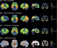
          Limbic age-related TDP-43 encephalopathy neuropathologic change (LATE-NC) is common in advanced age and can underlie a clinical presentation mimicking Alzheimer’s disease (AD). We studied whether an autopsy-derived fluorodeoxyglucose positron emission tomography (FDG-PET) signature of LATE-NC provides clinical utility for differential diagnosis of amnestic dementia patients.

          Methods:

          Ante mortem FDG-PET patterns from autopsy-confirmed LATE-NC ( N = 7) and AD ( N = 23) patients were used to stratify an independent cohort of clinically diagnosed AD dementia patients ( N = 242) based on individual FDG-PET profiles.

          Results:

          Autopsy-confirmed LATE-NC and AD groups showed markedly distinct temporo-limbic and temporo-parietal FDG-PET patterns, respectively. Clinically diagnosed AD dementia patients showing a LATE-NC–like FDG-PET pattern ( N = 25, 10%) were significantly older, showed less abnormal AD biomarker levels, lower APOE ε4, and higher TMEM106B risk allele load. Clinically, they exhibited a more memory-predominant profile and a generally slower disease course.

          Discussion:

          An autopsy-derived temporo-limbic FDG-PET signature identifies older amnestic patients whose clinical, genetic, and molecular biomarker features are consistent with underlying LATE-NC.

          Related collections

          Most cited references49

          • Record: found
          • Abstract: found
          • Article: found
          Is Open Access

          Calculating and reporting effect sizes to facilitate cumulative science: a practical primer for t-tests and ANOVAs

          Effect sizes are the most important outcome of empirical studies. Most articles on effect sizes highlight their importance to communicate the practical significance of results. For scientists themselves, effect sizes are most useful because they facilitate cumulative science. Effect sizes can be used to determine the sample size for follow-up studies, or examining effects across studies. This article aims to provide a practical primer on how to calculate and report effect sizes for t-tests and ANOVA's such that effect sizes can be used in a-priori power analyses and meta-analyses. Whereas many articles about effect sizes focus on between-subjects designs and address within-subjects designs only briefly, I provide a detailed overview of the similarities and differences between within- and between-subjects designs. I suggest that some research questions in experimental psychology examine inherently intra-individual effects, which makes effect sizes that incorporate the correlation between measures the best summary of the results. Finally, a supplementary spreadsheet is provided to make it as easy as possible for researchers to incorporate effect size calculations into their workflow.
            Bookmark
            • Record: found
            • Abstract: found
            • Article: not found

            National Institute on Aging-Alzheimer's Association guidelines for the neuropathologic assessment of Alzheimer's disease: a practical approach.

            We present a practical guide for the implementation of recently revised National Institute on Aging-Alzheimer's Association guidelines for the neuropathologic assessment of Alzheimer's disease (AD). Major revisions from previous consensus criteria are: (1) recognition that AD neuropathologic changes may occur in the apparent absence of cognitive impairment, (2) an "ABC" score for AD neuropathologic change that incorporates histopathologic assessments of amyloid β deposits (A), staging of neurofibrillary tangles (B), and scoring of neuritic plaques (C), and (3) more detailed approaches for assessing commonly co-morbid conditions such as Lewy body disease, vascular brain injury, hippocampal sclerosis, and TAR DNA binding protein (TDP)-43 immunoreactive inclusions. Recommendations also are made for the minimum sampling of brain, preferred staining methods with acceptable alternatives, reporting of results, and clinico-pathologic correlations.
              Bookmark
              • Record: found
              • Abstract: found
              • Article: not found

              When to use the Bonferroni correction.

              The Bonferroni correction adjusts probability (p) values because of the increased risk of a type I error when making multiple statistical tests. The routine use of this test has been criticised as deleterious to sound statistical judgment, testing the wrong hypothesis, and reducing the chance of a type I error but at the expense of a type II error; yet it remains popular in ophthalmic research. The purpose of this article was to survey the use of the Bonferroni correction in research articles published in three optometric journals, viz. Ophthalmic & Physiological Optics, Optometry & Vision Science, and Clinical & Experimental Optometry, and to provide advice to authors contemplating multiple testing.
                Bookmark

                Author and article information

                Journal
                101231978
                33173
                Alzheimers Dement
                Alzheimers Dement
                Alzheimer's & dementia : the journal of the Alzheimer's Association
                1552-5260
                1552-5279
                16 December 2022
                April 2023
                15 August 2022
                01 April 2024
                : 19
                : 4
                : 1234-1244
                Affiliations
                [1 ]Unidad de Trastornos del Movimiento, Servicio de Neurología y Neurofisiología Clínica, Instituto de Biomedicina de Sevilla, Hospital Universitario Virgen del Rocío/CSIC/Universidad de Sevilla, Seville, Spain
                [2 ]German Center for Neurodegenerative Diseases (DZNE), Rostock, Germany
                [3 ]Wallenberg Center for Molecular and Translational Medicine and Department of Psychiatry and Neurochemistry, University of Gothenburg, Gothenburg, Sweden
                [4 ]Charité – Universitätsmedizin Berlin, corporate member of Freie Universität Berlin and Humboldt-Universität zu Berlin, Department of Nuclear Medicine, Berlin, Germany
                [5 ]Indiana Alzheimer’s Disease Research Center and Department of Radiology and Imaging Sciences, Indiana University School of Medicine, Indianapolis, Indiana, USA
                [6 ]Sanders-Brown Center on Aging and Department of Pathology, University of Kentucky, Lexington, Kentucky, USA
                [7 ]Department of Neurodegenerative Disease, UCL Institute of Neurology, London, UK
                [8 ]Department of Diagnostic and Interventional Radiology and Nuclear Medicine, University Medical Center Hamburg-Eppendorf, Hamburg, Germany
                [9 ]Department of Psychosomatic Medicine, Rostock University Medical Center, Rostock, Germany
                Author notes
                Correspondence Michel J. Grothe, Unidad de Trastornos del Movimiento, Servicio de Neurología y, Neurofisiología Clínica, Instituto de Biomedicina de Sevilla, Hospital Universitario Virgen del Rocío/CSIC/Universidad de Sevilla, Avda.Manuel Siurot s/n, 41013 Seville, Spain. mgrothe@ 123456us.es
                Article
                NIHMS1851259
                10.1002/alz.12763
                9929029
                35971593
                1d227bdf-f5d3-45cd-91d7-d8780d28c4d1

                This is an open access article under the terms of the CreativeCommonsAttribution-NonCommercial License, which permits use, distribution and reproduction in any medium, provided the original work is properly cited and is not used for commercial purposes.

                History
                Categories
                Article

                amyloid,apolipoprotein e,autopsy,fluorodeoxyglucose positron emission tomography,hippocampal sclerosis,limbic age-related tdp-43 encephalopathy,tau,tdp-43,tmem106b

                Comments

                Comment on this article