0
views
0
recommends
+1 Recommend
0 collections
    0
    shares
      • Record: found
      • Abstract: found
      • Article: found
      Is Open Access

      AvaGen Genetic Testing versus Ocular Screening Assessments Including the Keratoconus Severity Score (KSS) and Randleman Ectasia Risk Score System (ERSS) in Refractive Surgery Candidates

      research-article

      Read this article at

      Bookmark
          There is no author summary for this article yet. Authors can add summaries to their articles on ScienceOpen to make them more accessible to a non-specialist audience.

          Abstract

          Purpose

          To determine whether the AvaGen (AG) Genetic Eye Test provided additional information for screening for the presence of keratoconus (KC) and assessing KC risk in refractive surgery candidates, as compared to the Keratoconus Severity Score (KSS) and Randleman Ectasia Risk Score System (ERSS).

          Methods

          This retrospective study analyzed patients seeking refractive surgery at an eye clinic in the United States between January 2022 and July 2023. The inclusion criteria encompassed those with a family history of KC, positive KC indices, or both. Corneal evaluations and demographic information were recorded and analyzed. KSS and ERSS criteria were utilized to evaluate postoperative KC and ectasia risk, respectively. Patients were categorized on how the AG genetic test compared to KSS and ERSS criteria. Clinicians assessed topographic indices, criteria scoring, and AG testing to deliver a definitive surgical recommendation.

          Results

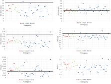
          Among the 19 patients evaluated for ectasia risk, AG testing showed lower KC risk than ocular screening in three patients (15.8%), equal risk in three patients (15.8%), and higher risk in 13 patients (68.4%). The mean AG scores were 45.7 ± 7.0, 49.0 ± 3.46, and 61 ± 13.0 for these respective categories. The most frequently identified KC risk genes were ADAMTS18, COL2A1, and COL4A1. The AG test modified the physician’s recommendation for refractive surgery in nine cases (47.4%).

          Conclusion

          Despite the promising application of AG testing for assessing KC risk, further research and development are needed to enhance its applicability for screening refractive surgery candidates, in addition to standard ocular screening approaches.

          Related collections

          Most cited references34

          • Record: found
          • Abstract: found
          • Article: not found

          Risk assessment for ectasia after corneal refractive surgery.

          To analyze the epidemiologic features of ectasia after excimer laser corneal refractive surgery, to identify risk factors for its development, and to devise a screening strategy to minimize its occurrence. Retrospective comparative and case-control study. All cases of ectasia after excimer laser corneal refractive surgery published in the English language with adequate information available through December 2005, unpublished cases seeking treatment at the authors' institution from 1998 through 2005, and a contemporaneous control group who underwent uneventful LASIK and experienced a normal postoperative course. Evaluation of preoperative characteristics, including patient age, gender, spherical equivalent refraction, pachymetry, and topographic patterns; perioperative characteristics, including type of surgery performed, flap thickness, ablation depth, and residual stromal bed (RSB) thickness; and postoperative characteristics including time to onset of ectasia. Development of postoperative corneal ectasia. There were 171 ectasia cases, including 158 published cases and 13 unpublished cases evaluated at the authors' institution. Ectasia occurred after LASIK in 164 cases (95.9%) and after photorefractive keratectomy (PRK) in 7 cases (4.1%). Compared with controls, more ectasia cases had abnormal preoperative topographies (35.7% vs. 0%; P<1.0x10(-15)), were significantly younger (34.4 vs. 40.0 years; P<1.0x10(-7)), were more myopic (-8.53 vs. -5.09 diopters; P<1.0x10(-7)), had thinner corneas before surgery (521.0 vs. 546.5 microm; P<1.0x10(-7)), and had less RSB thickness (256.3 vs. 317.3 microm; P<1.0x10(-10)). Based on subgroup logistic regression analysis, abnormal topography was the most significant factor that discriminated cases from controls, followed by RSB thickness, age, and preoperative corneal thickness, in that order. A risk factor stratification scale was created, taking all recognized risk factors into account in a weighted fashion. This model had a specificity of 91% and a sensitivity of 96% in this series. A quantitative method can be used to identify eyes at risk for developing ectasia after LASIK that, if validated, represents a significant improvement over current screening strategies.
            Bookmark
            • Record: found
            • Abstract: found
            • Article: found
            Is Open Access

            The Genetic and Environmental Factors for Keratoconus

            Keratoconus (KC) is the most common cornea ectatic disorder. It is characterized by a cone-shaped thin cornea leading to myopia, irregular astigmatism, and vision impairment. It affects all ethnic groups and both genders. Both environmental and genetic factors may contribute to its pathogenesis. This review is to summarize the current research development in KC epidemiology and genetic etiology. Environmental factors include but are not limited to eye rubbing, atopy, sun exposure, and geography. Genetic discoveries have been reviewed with evidence from family-based linkage analysis and fine mapping in linkage region, genome-wide association studies, and candidate genes analyses. A number of genes have been discovered at a relatively rapid pace. The detailed molecular mechanism underlying KC pathogenesis will significantly advance our understanding of KC and promote the development of potential therapies.
              Bookmark
              • Record: found
              • Abstract: found
              • Article: not found

              The Prevalence and Risk Factors for Keratoconus: A Systematic Review and Meta-Analysis

              This study was conducted to determine the prevalence and risk factors for keratoconus worldwide.
                Bookmark

                Author and article information

                Journal
                Clin Ophthalmol
                Clin Ophthalmol
                opth
                Clinical Ophthalmology (Auckland, N.Z.)
                Dove
                1177-5467
                1177-5483
                07 May 2024
                2024
                : 18
                : 1245-1255
                Affiliations
                [1 ]Hoopes Vision Research Center, Hoopes Vision , Draper, UT, USA
                [2 ]John A. Moran Eye Center, University of Utah School of Medicine , Salt Lake City, UT, USA
                [3 ]Utah Lions Eye Bank , Murray, UT, USA
                [4 ]University of Louisville School of Medicine , Louisville, KY, USA
                [5 ]University of Arizona College of Medicine-Phoenix , Phoenix, AZ, USA
                [6 ]Pacific Northwest University of Health Sciences , Yakima, WA, USA
                [7 ]University of Utah School of Medicine , Salt Lake City, UT, USA
                Author notes
                Correspondence: Majid Moshirfar, Hoopes Vision Research Center, 11820 S. State St. #200, Draper, UT, 84020, USA, Tel +1 801-568-0200, Fax +1 801-563-0200, Email cornea2020@me.com
                Author information
                http://orcid.org/0000-0003-1024-6250
                http://orcid.org/0000-0002-2567-8331
                Article
                452128
                10.2147/OPTH.S452128
                11088376
                38737593
                1c9b61aa-3635-4355-b328-84df1bb77b31
                © 2024 Moshirfar et al.

                This work is published and licensed by Dove Medical Press Limited. The full terms of this license are available at https://www.dovepress.com/terms.php and incorporate the Creative Commons Attribution – Non Commercial (unported, v3.0) License ( http://creativecommons.org/licenses/by-nc/3.0/). By accessing the work you hereby accept the Terms. Non-commercial uses of the work are permitted without any further permission from Dove Medical Press Limited, provided the work is properly attributed. For permission for commercial use of this work, please see paragraphs 4.2 and 5 of our Terms ( https://www.dovepress.com/terms.php).

                History
                : 15 December 2023
                : 15 April 2024
                Page count
                Figures: 6, Tables: 3, References: 38, Pages: 11
                Categories
                Original Research

                Ophthalmology & Optometry
                avagen,keratoconus,severity,corneal topography,randleman,ectasia risk score system,keratoconus severity score

                Comments

                Comment on this article