3
views
0
recommends
+1 Recommend
1 collections
    0
    shares
      • Record: found
      • Abstract: found
      • Article: found
      Is Open Access

      Inverse folding of protein complexes with a structure-informed language model enables unsupervised antibody evolution

      Preprint
      research-article

      Read this article at

      Bookmark
          There is no author summary for this article yet. Authors can add summaries to their articles on ScienceOpen to make them more accessible to a non-specialist audience.

          Abstract

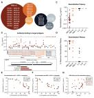
          Large language models trained on sequence information alone are capable of learning high level principles of protein design. However, beyond sequence, the three-dimensional structures of proteins determine their specific function, activity, and evolvability. Here we show that a general protein language model augmented with protein structure backbone coordinates and trained on the inverse folding problem can guide evolution for diverse proteins without needing to explicitly model individual functional tasks. We demonstrate inverse folding to be an effective unsupervised, structure-based sequence optimization strategy that also generalizes to multimeric complexes by implicitly learning features of binding and amino acid epistasis. Using this approach, we screened ~30 variants of two therapeutic clinical antibodies used to treat SARS-CoV-2 infection and achieved up to 26-fold improvement in neutralization and 37-fold improvement in affinity against antibody-escaped viral variants-of-concern BQ.1.1 and XBB.1.5, respectively. In addition to substantial overall improvements in protein function, we find inverse folding performs with leading experimental success rates among other reported machine learning-guided directed evolution methods, without requiring any task-specific training data.

          Related collections

          Most cited references63

          • Record: found
          • Abstract: found
          • Article: found
          Is Open Access

          Highly accurate protein structure prediction with AlphaFold

          Proteins are essential to life, and understanding their structure can facilitate a mechanistic understanding of their function. Through an enormous experimental effort 1 – 4 , the structures of around 100,000 unique proteins have been determined 5 , but this represents a small fraction of the billions of known protein sequences 6 , 7 . Structural coverage is bottlenecked by the months to years of painstaking effort required to determine a single protein structure. Accurate computational approaches are needed to address this gap and to enable large-scale structural bioinformatics. Predicting the three-dimensional structure that a protein will adopt based solely on its amino acid sequence—the structure prediction component of the ‘protein folding problem’ 8 —has been an important open research problem for more than 50 years 9 . Despite recent progress 10 – 14 , existing methods fall far short of atomic accuracy, especially when no homologous structure is available. Here we provide the first computational method that can regularly predict protein structures with atomic accuracy even in cases in which no similar structure is known. We validated an entirely redesigned version of our neural network-based model, AlphaFold, in the challenging 14th Critical Assessment of protein Structure Prediction (CASP14) 15 , demonstrating accuracy competitive with experimental structures in a majority of cases and greatly outperforming other methods. Underpinning the latest version of AlphaFold is a novel machine learning approach that incorporates physical and biological knowledge about protein structure, leveraging multi-sequence alignments, into the design of the deep learning algorithm. AlphaFold predicts protein structures with an accuracy competitive with experimental structures in the majority of cases using a novel deep learning architecture.
            Bookmark
            • Record: found
            • Abstract: found
            • Article: found
            Is Open Access

            Isolation of potent SARS-CoV-2 neutralizing antibodies and protection from disease in a small animal model

            Countermeasures to prevent and treat COVID-19 are a global health priority. We enrolled a cohort of SARS-CoV-2-recovered participants, developed neutralization assays to interrogate antibody responses, adapted our high-throughput antibody generation pipeline to rapidly screen over 1800 antibodies, and established an animal model to test protection. We isolated potent neutralizing antibodies (nAbs) to two epitopes on the receptor binding domain (RBD) and to distinct non-RBD epitopes on the spike (S) protein. We showed that passive transfer of a nAb provides protection against disease in high-dose SARS-CoV-2 challenge in Syrian hamsters, as revealed by maintained weight and low lung viral titers in treated animals. The study suggests a role for nAbs in prophylaxis, and potentially therapy, of COVID-19. The nAbs define protective epitopes to guide vaccine design.
              Bookmark
              • Record: found
              • Abstract: found
              • Article: found
              Is Open Access

              BA.2.12.1, BA.4 and BA.5 escape antibodies elicited by Omicron infection

              Severe acute respiratory syndrome coronavirus 2 (SARS-CoV-2) Omicron sublineages BA.2.12.1, BA.4 and BA.5 exhibit higher transmissibility than the BA.2 lineage 1 . The receptor binding and immune-evasion capability of these recently emerged variants require immediate investigation. Here, coupled with structural comparisons of the spike proteins, we show that BA.2.12.1, BA.4 and BA.5 (BA.4 and BA.5 are hereafter referred collectively to as BA.4/BA.5) exhibit similar binding affinities to BA.2 for the angiotensin-converting enzyme 2 (ACE2) receptor. Of note, BA.2.12.1 and BA.4/BA.5 display increased evasion of neutralizing antibodies compared with BA.2 against plasma from triple-vaccinated individuals or from individuals who developed a BA.1 infection after vaccination. To delineate the underlying antibody-evasion mechanism, we determined the escape mutation profiles 2 , epitope distribution 3 and Omicron-neutralization efficiency of 1,640 neutralizing antibodies directed against the receptor-binding domain of the viral spike protein, including 614 antibodies isolated from people who had recovered from BA.1 infection. BA.1 infection after vaccination predominantly recalls humoral immune memory directed against ancestral (hereafter referred to as wild-type (WT)) SARS-CoV-2 spike protein. The resulting elicited antibodies could neutralize both WT SARS-CoV-2 and BA.1 and are enriched on epitopes on spike that do not bind ACE2. However, most of these cross-reactive neutralizing antibodies are evaded by spike mutants L452Q, L452R and F486V. BA.1 infection can also induce new clones of BA.1-specific antibodies that potently neutralize BA.1. Nevertheless, these neutralizing antibodies are largely evaded by BA.2 and BA.4/BA.5 owing to D405N and F486V mutations, and react weakly to pre-Omicron variants, exhibiting narrow neutralization breadths. The therapeutic neutralizing antibodies bebtelovimab 4 and cilgavimab 5 can effectively neutralize BA.2.12.1 and BA.4/BA.5, whereas the S371F, D405N and R408S mutations undermine most broadly sarbecovirus-neutralizing antibodies. Together, our results indicate that Omicron may evolve mutations to evade the humoral immunity elicited by BA.1 infection, suggesting that BA.1-derived vaccine boosters may not achieve broad-spectrum protection against new Omicron variants.
                Bookmark

                Author and article information

                Journal
                bioRxiv
                BIORXIV
                bioRxiv
                Cold Spring Harbor Laboratory
                21 December 2023
                : 2023.12.19.572475
                Affiliations
                [1 ]Stanford Biophysics Program, Stanford University School of Medicine, Stanford, CA 94305, USA
                [2 ]Stanford Medical Scientist Training Program, Stanford University School of Medicine, Stanford CA 94305, USA
                [3 ]Sarafan ChEM-H, Stanford University, Stanford, CA 94305, USA
                [4 ]Department of Biochemistry, Stanford University School of Medicine, Stanford, CA 94305, USA
                [5 ]Chan Zuckerberg Biohub, San Francisco, CA 94158, USA
                Author notes

                Author contributions

                Conceptualization, methodology, interpretation: V.R.S., B.L.H., P.S.K.; Computational experiments and software development: V.R.S.; Antibody and antigen cloning, expression, and purification: V.R.S., T.U.J.B.; Lentivirus production and pseudovirus neutralization: T.U.J.B; Binding assays: V.R.S.; Writing (original draft): V.R.S with assistance from B.L.H and P.S.K.; Writing (final draft): all authors

                [* ]Correspondence: B.L.H. ( brianhie@ 123456stanford.edu ), P.S.K. ( kimpeter@ 123456stanford.edu )
                Author information
                http://orcid.org/0000-0003-4443-9229
                http://orcid.org/0000-0002-7462-2537
                http://orcid.org/0000-0003-3224-8142
                http://orcid.org/0000-0001-6503-4541
                Article
                10.1101/2023.12.19.572475
                10769282
                38187780
                1c14a3b3-98b0-4e0e-af9d-9c011c7d5f31

                This work is licensed under a Creative Commons Attribution-NonCommercial-NoDerivatives 4.0 International License, which allows reusers to copy and distribute the material in any medium or format in unadapted form only, for noncommercial purposes only, and only so long as attribution is given to the creator.

                History
                Categories
                Article

                Comments

                Comment on this article